All SubjectsAnatomy (167)Anesthesiology (9)Biochemistry (108)Community Medicine (87)Dental (8)Dermatology (19)ENT (30)Forensic Medicine (62)General Medicine (3)Internal Medicine (136)Microbiology (101)Obstetrics and Gynecology (65)Ophthalmology (60)Orthopaedics (33)Pathology (107)Pediatrics (37)Pharmacology (123)Physiology (116)Psychiatry (2)Psychiatry (38)Radiology (25)Surgery (81)
Q11
What is the immediate emergency treatment for carbon monoxide (CO) poisoning?
Q12
Which of the following is not a feature of Poststreptococcal Glomerulonephritis (PSGN)?
Q13
Which type of thyroid cancer is associated with primary hyperparathyroidism and phaeochromocytoma?
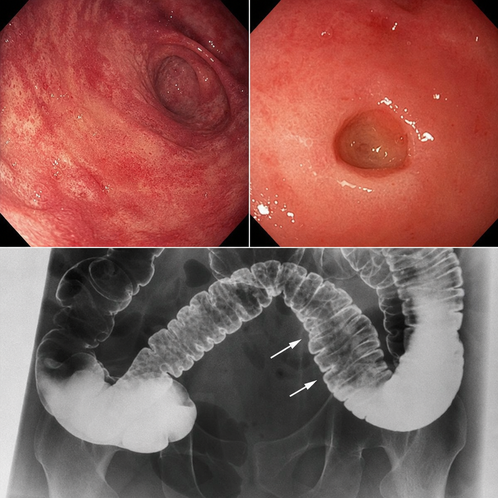
Q14
A patient presents with skin involvement and collar stud ulceration in the intestine observed on radiography. What is the most likely diagnosis?
Q15
In which condition is the Doll's Eye Reflex tested?
Q16
Which subtype of Acute Myeloid Leukemia (AML) is associated with the best prognosis?
Q17
In which condition is Serum Amyloid Associated (SAA) protein most commonly found?
Q18
Shrinking Lung Syndrome is seen in:
Q19
What is the primary effect of beta blockers in the management of thyroid storm?
Q20
Extremities are warm in which type of shock