All (126)Anatomy (11)Anesthesiology (3)Biochemistry (11)Community Medicine (2)Dental (1)Dermatology (1)ENT (2)Forensic Medicine (2)General Medicine (1)Internal Medicine (17)Microbiology (10)Obstetrics and Gynecology (5)Ophthalmology (6)Orthopaedics (2)Pathology (9)Pediatrics (11)Pharmacology (6)Physiology (6)Psychiatry (6)Radiology (5)Surgery (9)
Q11
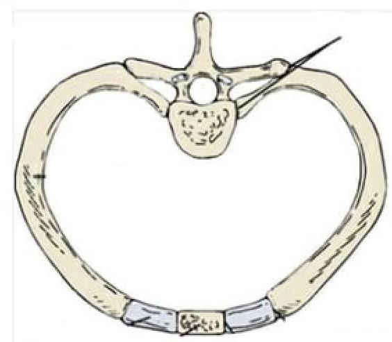
Identify the type of joint in the image provided.
Q12
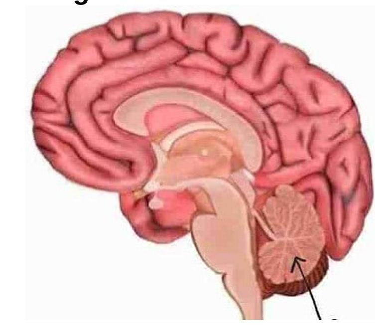
Identify the marked structure in the image.