INI-CET 2024 — Anatomy
9 Previous Year Questions with Answers & Explanations
The thoracic duct crosses from the right to the left at the level of
Identify the marked structure in the image.
Identify the type of joint in the image provided.
Which of the following statements is true regarding the anatomy of the breast?
At which vertebral level is the ischial spine located?
Which of the following is the PRIMARY action of the superior oblique muscle?
Substantia nigra is connected to which part of the basal ganglia?
Which structure is NOT present in the floor of the inferior horn of the lateral ventricle?
The bone matrix has the following crystals -
INI-CET 2024 - Anatomy INI-CET Practice Questions and MCQs
Question 1: The thoracic duct crosses from the right to the left at the level of
- A. T12 vertebra
- B. T2 vertebra
- C. T4-T5 vertebra (Correct Answer)
- D. T6 vertebra
Explanation: ***T4-T5 vertebra*** - The **thoracic duct** crosses from the right to the left side of the vertebral column at the level of the **T4-T5 vertebrae**, specifically just above the root of the left lung. - This crossover is an important anatomical landmark as it signifies the duct's ascent towards the neck to drain into the left subclavian vein. *T12 vertebra* - The **thoracic duct** originates from the **cisterna chyli** at the level of the L1 or L2 vertebra and ascends into the thorax at or below the T12 vertebra, it does not cross over at this level. - This level primarily marks its entry into the thoracic cavity, not its main crossover point. *T6 vertebra* - While the **thoracic duct** is present in the thorax at this level, it does not undergo its characteristic crossover from right to left at the T6 vertebra. - The duct continues its ascent along the right side of the vertebral column before moving across. *T2 vertebra* - By the level of the T2 vertebra, the **thoracic duct** has already crossed to the left side of the vertebral column and is ascending towards its termination in the neck. - The crossover event occurs more inferiorly, at the T4-T5 level.
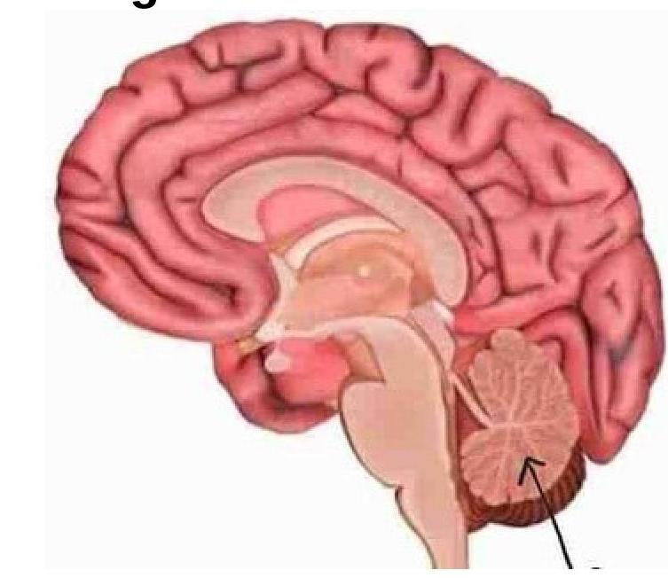
Question 2: Identify the marked structure in the image.
- A. Cerebrum
- B. Brain stem
- C. Corpus callosum
- D. Cerebellum (Correct Answer)
Explanation: ***Cerebellum*** - The image points to the distinct, posterior inferior structure of the brain, characterized by its **folia** and arbour-vitae-like internal structure, which is the cerebellum. - The cerebellum is primarily involved in **motor control**, including coordination, precision, and accurate timing. *Cerebrum* - The cerebrum is the **largest part of the brain**, located superiorly, responsible for higher functions like thought, voluntary movement, and sensory processing. - It consists of two hemispheres connected by the corpus callosum and is characterized by its **gyri** and **sulci**. *Brain stem* - The brain stem is located inferior to the cerebrum and anterior to the cerebellum, connecting the cerebrum and cerebellum to the **spinal cord**. - It controls vital functions such as **breathing**, heart rate, and sleep, and is composed of the midbrain, pons, and medulla oblongata. *Corpus callosum* - The corpus callosum is a large, C-shaped nerve fiber bundle located deep within the brain, under the cerebral cortex. - Its primary function is to **connect the two cerebral hemispheres**, facilitating communication between them.
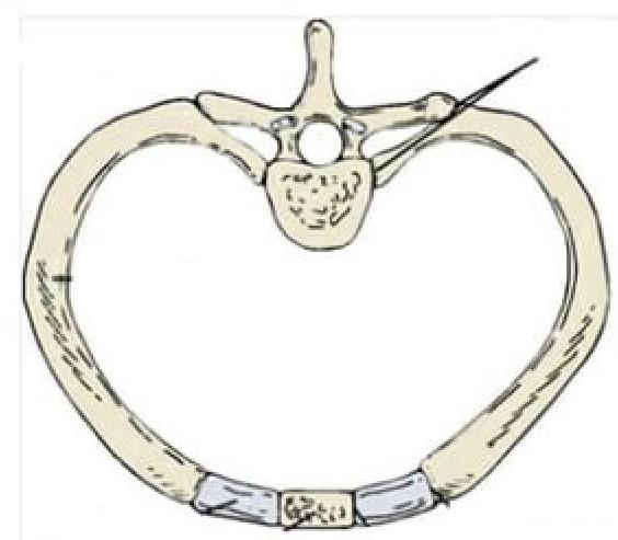
Question 3: Identify the type of joint in the image provided.
- A. Syndesmosis
- B. Synarthrosis
- C. Synovial joint (Correct Answer)
- D. Symphysis
Explanation: ***Synovial joint*** - The image depicts a **costovertebral joint**, which connects a rib to a thoracic vertebra. These joints are **diarthrotic**, meaning they are freely movable, characteristic of synovial joints. - Synovial joints are characterized by the presence of a **synovial cavity**, articular cartilage, an articular capsule, and synovial fluid, allowing for a wide range of motion. *Syndesmosis* - A syndesmosis is a type of **fibrous joint** where two bones are joined by a ligament or a membrane, allowing for very limited movement, such as the distal tibiofibular joint. - This definition does not match the image, which shows a joint designed for movement between the rib and vertebra. *Synarthrosis* - Synarthrosis is a classification for **immovable joints**, such as sutures in the skull. - The costovertebral joints, as shown, allow for movement during respiration and are therefore not synarthrotic. *Symphysis* - A symphysis is a type of **cartilaginous joint** where bones are joined by **fibrocartilage**, allowing for slight movement. Examples include the pubic symphysis or intervertebral discs. - The costovertebral joint shown in the image is a synovial articulation, not a cartilaginous joint.
Question 4: Which of the following statements is true regarding the anatomy of the breast?
- A. The superior medial quadrant has more tissue
- B. The nipple is located at the level of the fourth intercostal space in most women
- C. There are 15-20 lobules present
- D. The axillary tail of Spence crosses the anterior axillary fold (Correct Answer)
Explanation: ***The axillary tail of Spence crosses the anterior axillary fold*** - The **axillary tail of Spence** is an extension of breast glandular tissue that passes superolaterally from the main breast body and often **penetrates the deep fascia** in the axilla [3]. - Its presence crossing the **anterior axillary fold** is relevant for physical examination and surgical considerations, as it can be a site for breast pathologies. *The superior medial quadrant has more tissue* - The **superior lateral quadrant** of the breast typically contains the **most glandular tissue** and lymphatics, making it the most common site for breast cancers. - This anatomical distribution is crucial for understanding the **etiology and metastasis** of breast malignancies. *The nipple is located at the level of the fourth intercostal space in most women* - The **nipple** typically lies at the level of the **fourth rib (not intercostal space)** in nulliparous women, but its position can vary significantly based on individual factors like breast size, age, and parity [1]. - Topographical landmarks such as the **midclavicular line** are often used for more consistent localization. *There are 15-20 lobules present* - Each breast typically contains **15-20 lobes**, not lobules, arranged radially around the nipple [1]. - Each **lobe** consists of numerous smaller **lobules**, which are the functional units of milk production, draining into ducts that converge at the nipple [2].
Question 5: At which vertebral level is the ischial spine located?
- A. L4
- B. S2 (Correct Answer)
- C. S3-S5
- D. Coccygeal region
Explanation: ***S2*** - The **ischial spine** is a bony projection located on the posterior border of the ischium [1]. - It is anatomically located at the vertebral level of **S2**, which is a crucial landmark especially in obstetrics for assessing the station of the fetal head during labor [1]. *L4* - The **L4 vertebral level** is generally associated with the **iliac crests** and is a common site for lumbar punctures. - It lies significantly superior to the ischial spine and is not a relevant landmark for its location. *S3-S5* - The vertebral levels **S3 to S5** primarily contribute to the formation of the lower sacrum and the coccyx. - While they are inferior to S2, they are not directly associated with the precise anatomical level of the ischial spine. *Coccygeal region* - The **coccygeal region** consists of the fused coccygeal vertebrae, forming the tailbone. - This region is located inferior to the sacrum and is distinct from the level of the ischial spine, which is situated higher on the pelvis.
Question 6: Which of the following is the PRIMARY action of the superior oblique muscle?
- A. Adduction
- B. Intorsion (Correct Answer)
- C. Abduction
- D. Depression
Explanation: ***Intorsion*** - The **primary action** of the **superior oblique muscle** is **intorsion** (internal rotation), which means rotating the top of the eyeball medially (towards the nose). - This action helps to counteract the **extorsion** caused by the inferior oblique muscle and stabilize the visual field during head tilt. *Depression* - While the superior oblique muscle does contribute to **depression** (moving the eye downwards), this is a **secondary action**, particularly when the eye is in **abduction** [1]. - The **inferior rectus muscle** is the primary depressor of the eye [1]. *Abduction* - The superior oblique has a minor **tertiary action** of **abduction** (moving the eye away from the midline) [1]. - However, the **lateral rectus muscle** is the primary abductor of the eye [1]. *Adduction* - **Adduction** (moving the eye towards the midline) is primarily performed by the **medial rectus muscle** [1]. - The superior oblique muscle does **NOT** contribute to adduction; this is not one of its actions [1].
Question 7: Substantia nigra is connected to which part of the basal ganglia?
- A. Thalamus
- B. Pallidum
- C. Striatum (Correct Answer)
- D. Subthalamic nucleus
Explanation: ***Striatum*** - The **substantia nigra pars compacta (SNc)** provides **dopaminergic input** to the striatum via the **nigrostriatal pathway**, which is crucial for motor control [1]. - This connection establishes the direct and indirect pathways of the basal ganglia, modulating **movement initiation** and **inhibition** [1]. *Thalamus* - The thalamus acts as a **relay station** for information leaving the basal ganglia, but it is not directly connected to the substantia nigra as a primary input or output structure within the basal ganglia circuitry [1]. - The basal ganglia influence the thalamus, which then projects to the **motor cortex**, but the direct connection from substantia nigra is to the striatum. *Pallidum* - The **pallidum (globus pallidus)** receives input from the striatum and projects to the thalamus, but it is not directly connected to the substantia nigra as the **primary recipient** of nigral efferents [1]. - While it's part of the basal ganglia, the substantia nigra's main direct projection is to the **striatum**. *Subthalamic nucleus* - The **subthalamic nucleus (STN)** is an excitatory component of the basal ganglia that receives input from the cortex and projects to the globus pallidus. - While there are some indirect connections, the STN is not the primary target of the **nigrostriatal dopaminergic projections** from the substantia nigra [1].
Question 8: Which structure is NOT present in the floor of the inferior horn of the lateral ventricle?
- A. Tail of the caudate nucleus (Correct Answer)
- B. Fimbria
- C. Hippocampus
- D. Collateral eminence
Explanation: ***Tail of the caudate nucleus*** - The **tail of the caudate nucleus** is located in the **roof** of the inferior horn of the lateral ventricle, not in the floor. - It courses along the lateral aspect of the inferior horn, terminating in the **amygdaloid body** [1]. *Fimbria* - The **fimbria** is a prominent white matter bundle that forms part of the **floor** of the inferior horn of the lateral ventricle. - It consists of efferent fibers from the hippocampus, converging to form the **crus of the fornix**. *Hippocampus* - The **hippocampus** is a major structure in the **floor** of the inferior horn of the lateral ventricle, forming a distinctive bulge [1]. - It plays a critical role in **memory formation** and extends throughout the length of the inferior horn [1]. *Collateral eminence* - The **collateral eminence** is an elevation in the **floor** of the inferior horn, lateral to the hippocampus. - It is formed by the indentation of the collateral sulcus on the inferior surface of the temporal lobe.
Question 9: The bone matrix has the following crystals -
- A. Calcium pyrophosphate
- B. Calcium hydroxyapatite (Correct Answer)
- C. Calcium phosphate
- D. Calcium sulphate
Explanation: ***Calcium hydroxyapatite*** - The primary mineral component of bone matrix is **calcium hydroxyapatite**, which gives bone its rigidity and strength [1]. - These crystals are formed from **calcium and phosphate ions** arranged in a specific crystalline structure within the collagen fibers [1]. *Calcium pyrophosphate* - **Calcium pyrophosphate dihydrate (CPPD)** crystals are associated with **pseudogout**, a condition causing joint inflammation, not the normal bone matrix [1]. - They are found in articular cartilage and synovial fluid, not as a structural component of healthy bone. *Calcium phosphate* - While hydroxyapatite is a form of **calcium phosphate**, simply "calcium phosphate" is too general and does not specify the exact crystalline structure found in bone [1]. - Many calcium phosphate compounds exist, but **hydroxyapatite** is the specific and most abundant one in bone [1]. *Calcium sulphate* - **Calcium sulfate** is not a naturally occurring mineral component of the bone matrix in vertebrates. - It is sometimes used in medical applications as a **bone graft substitute** or a drug delivery system, but not as an endogenous component.