FMGE 2017 — Physiology
5 Previous Year Questions with Answers & Explanations
Which of the following ion plays a role in exocytosis?
In which of the following conditions is blood osmolality increased?
The PR interval in ECG denotes?
Which of these is true about SGLT1?
In the process shown below stretch stimulus is mediated by which of the following receptors?
FMGE 2017 - Physiology FMGE Practice Questions and MCQs
Question 1: Which of the following ion plays a role in exocytosis?
- A. Potassium
- B. Sodium
- C. Calcium (Correct Answer)
- D. Magnesium
Explanation: ***Calcium*** - **Calcium ions** are crucial for initiating the fusion of **secretory vesicles** with the plasma membrane during **exocytosis**. - An increase in intracellular calcium concentration, often due to an influx from outside the cell, triggers the release of neurotransmitters, hormones, and other substances. *Potassium* - **Potassium ions** are primarily involved in maintaining the **resting membrane potential** and repolarization during action potentials. - While essential for neuronal function, they do not directly trigger the **vesicle fusion** step of exocytosis. *Sodium* - **Sodium ions** are vital for depolarizing the membrane and initiating **action potentials**, as well as for certain co-transport mechanisms. - However, they do not directly bind to proteins involved in **exocytosis** to trigger the release mechanism. *Magnesium* - **Magnesium ions** serve as **cofactors** for many enzymes, including ATPases, and play a role in stabilizing nucleic acids and proteins. - While important for overall cellular function, magnesium does not directly initiate or regulate the **fusion events** of exocytosis.
Question 2: In which of the following conditions is blood osmolality increased?
- A. SIADH
- B. Psychogenic polydipsia
- C. Diarrhea (Correct Answer)
- D. Cerebral toxoplasmosis
Explanation: ***Diarrhea*** - Diarrhea leads to a significant loss of **water and electrolytes** from the body, primarily from the extracellular fluid compartment. - This imbalance causes **hemoconcentration** and an increase in the concentration of solutes in the blood, thereby raising blood osmolality. *SIADH* - **Syndrome of Inappropriate Antidiuretic Hormone (SIADH)** is characterized by excessive secretion of ADH, leading to **dilutional hyponatremia**. - The excess water retention dilutes the blood, resulting in **decreased serum osmolality**. *Psychogenic polydipsia* - This condition involves excessive water intake due to psychological factors, which causes **dilution of body fluids**. - The increased water volume without a proportional increase in solutes leads to **decreased plasma osmolality**. *Cerebral toxoplasmosis* - **Cerebral toxoplasmosis** is an opportunistic infection of the brain, typically seen in immunocompromised individuals. - It primarily causes neurological symptoms and **does not directly impact blood osmolality** unless complicated by other factors like dehydration or SIADH (which is not a primary effect).
Question 3: The PR interval in ECG denotes?
- A. Ventricular depolarization and ventricular repolarization
- B. Atrial depolarization with atrial repolarization
- C. Atrial depolarization with A - V conduction (Correct Answer)
- D. Atrial depolarization only
Explanation: ***Atrial depolarization with A - V conduction*** * The **PR interval** reflects the time from the beginning of **atrial depolarization** (P wave) to the beginning of **ventricular depolarization** (QRS complex). * It represents the time taken for the electrical impulse to travel through the **atria** and the **AV node** to the ventricles. *Ventricular depolarization and ventricular repolarization* * **Ventricular depolarization** is represented by the **QRS complex**, and **ventricular repolarization** is represented by the **T wave**. * The PR interval occurs before the QRS complex, not during ventricular depolarization or repolarization. *Atrial depolarization with atrial repolarization* * **Atrial depolarization** is represented by the **P wave**. * **Atrial repolarization** typically occurs simultaneously with **ventricular depolarization** (QRS complex) and is often obscured by it. The PR interval includes the P wave but extends beyond it. *Atrial depolarization only* * **Atrial depolarization** is solely represented by the **P wave**. * The PR interval is a longer duration that includes the P wave and the subsequent delay in the **AV node**.
Question 4: Which of these is true about SGLT1?
- A. Secondary active transport of glucose in prostate
- B. Secondary active transport of glucose in brain
- C. Secondary active transport of glucose in intestine (Correct Answer)
- D. Secondary active transport of glucose in rods and cones
Explanation: ***Secondary active transport of glucose in intestine*** - **SGLT1** is the primary transporter responsible for **glucose and galactose absorption** from the lumen of the small intestine into the enterocytes. - It uses the electrochemical gradient of **sodium** to co-transport glucose against its concentration gradient, classifying it as **secondary active transport**. *Secondary active transport of glucose in prostate* - While glucose is vital for prostate metabolism, its transport predominantly involves **GLUTs** (e.g., GLUT1), not SGLT1. - SGLT1 is generally not found in significant amounts in the prostate. *Secondary active transport of glucose in brain* - Glucose transport across the **blood-brain barrier** and into brain cells is primarily mediated by **GLUT1** and other GLUT transporters, which are **facilitated diffusers**, not SGLT1. - SGLT1 has a very limited role, if any, in normal brain glucose uptake. *Secondary active transport of glucose in rods and cones* - Retinal photoreceptors (rods and cones) indeed rely on glucose, but its uptake is mainly via **GLUT1** and other GLUT family members. - **SGLT1** is not a significant transporter for glucose in these cells.
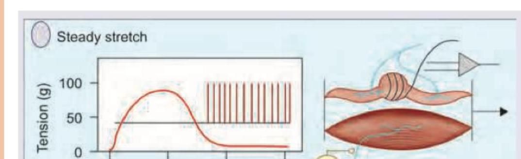
Question 5: In the process shown below stretch stimulus is mediated by which of the following receptors?
- A. Merkel's disc
- B. Meissner's corpuscle
- C. Pacinian corpuscle
- D. Muscle spindle (Correct Answer)
Explanation: ***Muscle spindle*** - The image depicts a **muscle**, and the "Steady stretch" stimulus clearly shows an increase in muscle tension followed by sustained neural firing, characteristic of a **stretch reflex**. - **Muscle spindles** are proprioceptors located within skeletal muscles that detect changes in muscle length and the rate of change of length, playing a crucial role in the stretch reflex. *Merkel's disc* - **Merkel's discs** are mechanoreceptors located in the basal layer of the epidermis, primarily responsible for detecting sustained light touch and pressure. - They are not involved in sensing muscle stretch. *Meissner's corpuscle* - **Meissner's corpuscles** are rapidly adapting mechanoreceptors found in the dermal papillae, specialized for detecting light touch and low-frequency vibration. - They are cutaneous receptors and do not mediate muscle stretch. *Pacinian corpuscle* - **Pacinian corpuscles** are rapidly adapting mechanoreceptors located deep in the dermis and subcutaneous tissue, sensitive to deep pressure and high-frequency vibration. - They are not responsible for detecting muscle stretch.