
Forensic Anthropology

On this page
🔍 Skeletal Foundations - Bone Blueprint Basics
When skeletal remains arrive at the morgue, they carry a silent biography written in bone-age etched in growth plates, sex coded in pelvic architecture, ancestry hinted in cranial contours, and stature calculated from limb proportions. You'll learn to read this osseous language systematically, moving from foundational anatomy through the forensic markers that transform fragments into identity. By mastering these principles, you'll understand how anthropologists reconstruct who someone was, bridging the gap between anonymous remains and meaningful answers for families, investigators, and justice systems worldwide.
📌 Remember: COMPACT for bone composition - Calcium phosphate (65% mineral), Organic matrix (35% collagen), Marrow spaces, Periosteum covering, Arterial supply, Cortical and cancellous architecture, Trabecular patterns
Bone Composition and Structure
Bone tissue consists of 65% inorganic minerals (primarily hydroxyapatite crystals) and 35% organic matrix (mainly Type I collagen). This composition creates the unique preservation characteristics that make skeletal remains valuable for forensic analysis, with mineral content providing durability while organic components retain microscopic structural details.
- Cortical Bone Architecture
- Dense outer layer comprising 80% of skeletal mass
- Haversian systems with 4-20 lamellae per osteon
- Porosity increases 0.5-2% per decade after age 30
- Forensic significance: Age estimation through histomorphometry
- Clinical correlation: Bone density changes reflect metabolic status
- Cancellous Bone Organization
- Trabecular network with 15-25% bone volume fraction
- Surface area 10x greater than cortical bone
- Remodeling rate 8x faster than cortical tissue
- Forensic application: Rapid response to pathological conditions
- Preservation factor: More susceptible to taphonomic changes

⭐ Clinical Pearl: Bone mineral density decreases 1-2% annually after menopause in females, creating sex-specific aging patterns detectable through microscopic analysis with 85% accuracy in age estimation.
Skeletal Classification Systems
Forensic anthropologists utilize multiple classification systems to organize skeletal analysis systematically. Bones are categorized by shape, development, and anatomical location, each category providing specific forensic information.
| Bone Type | Examples | Forensic Significance | Key Measurements | Accuracy Rate |
|---|---|---|---|---|
| Long Bones | Femur, Tibia, Humerus | Stature estimation | Maximum length ±2mm | 95% for height |
| Flat Bones | Skull vault, Pelvis | Sex determination | Thickness 2-8mm | 90% for sex |
| Short Bones | Carpals, Tarsals | Age assessment | Fusion timing ±6 months | 85% for age |
| Irregular | Vertebrae, Sacrum | Pathology detection | Morphological variants | 80% for disease |
| Sesamoid | Patella, Pisiform | Individual variation | Presence/absence | 70% for ID |
Anatomical Variation and Population Differences
Skeletal variation reflects genetic, environmental, and biomechanical influences that create population-specific patterns essential for ancestry assessment. Understanding these variations prevents misidentification and enables accurate demographic profiling.
Loading diagram…
- Population-Specific Characteristics
- Cranial index varies 15-20% between populations
- Femoral curvature differs by 5-10 degrees ancestry-dependent
- Dental morphology shows 60+ discrete traits
- Asian populations: Shovel-shaped incisors (85% frequency)
- African populations: Three-rooted mandibular molars (20% frequency)
- European populations: Carabelli's cusp (60% frequency)
📌 Remember: ASIA for Asian cranial features - Anterior nasal spine reduced, Shovel-shaped incisors, Incisive foramen heart-shaped, Angle of nasal bones obtuse (>130 degrees)
Bone Development and Maturation
Skeletal development follows predictable patterns that enable precise age estimation, particularly in subadults. Understanding ossification sequences, epiphyseal fusion timing, and growth patterns provides the foundation for forensic age assessment.
- Ossification Centers and Timing
- Primary centers appear 6-8 weeks gestational age
- Secondary centers emerge birth to 20 years
- Fusion completion varies ±2 years from population means
- Clavicle: Latest fusion at 25-30 years
- Iliac crest: Fusion at 20-23 years
- Vertebral rings: Complete by 25 years
⭐ Clinical Pearl: The medial clavicular epiphysis remains unfused until 25-30 years, making it the most reliable indicator for distinguishing juveniles from adults in forensic contexts with 90% accuracy.
Understanding skeletal foundations enables forensic anthropologists to approach human remains systematically, extracting maximum biological information from fragmentary evidence. This knowledge forms the cornerstone for advanced analytical techniques in demographic assessment and individual identification.
🔍 Skeletal Foundations — Bone Blueprint Basics


🧬 Age & Ossification - Bony Age Almanac
Age estimation from skeletal remains relies on predictable developmental and degenerative changes that occur throughout life. These changes follow established chronological patterns, though individual variation and population differences must be considered. Forensic anthropologists utilize multiple age indicators simultaneously to achieve maximum accuracy, recognizing that precision decreases with advancing age. Under BNSS procedures, accurate age determination becomes critical for establishing jurisdiction and applicable legal provisions.
📌 Remember: FUSED for epiphyseal union sequence - Fibula head (16-18 years), Ulnar olecranon (14-16 years), Sacral segments (18-25 years), Elbow epicondyles (13-15 years), Distal radius (17-19 years)
Fetal and Infant Age Assessment
Fetal age estimation is based on long bone lengths, individual bone development, and developing dentition. Infant and child age estimation relies on dental and skeletal indicators, including dental development, dental eruption, diaphyseal dimensions, and the appearance and maturation of ossification centers. This accuracy stems from rapid, predictable growth rates and consistent ossification timing during early development, achieving precision within ±2 weeks for fetal remains and ±4 months for infants up to 2 years.
- Fetal Age Indicators (Gestational Weeks)
- Clavicle ossification: 5-6 weeks
- Mandible ossification: 6-7 weeks
- Maxilla and rib formation: 7-8 weeks
- Femur diaphyseal length: 7mm at 12 weeks, 35mm at 20 weeks
- Humerus diaphyseal length: 6mm at 12 weeks, 30mm at 20 weeks
- Infant Ossification Milestones
- Distal femoral epiphysis: 36-40 weeks gestational
- Proximal tibial epiphysis: birth to 3 months
- Cuboid ossification: birth to 6 months
- Crown-heel length correlation: ±1.5cm accuracy
- Dental development: First molar calcification at birth
⭐ Clinical Pearl: The distal femoral epiphysis appears 2-4 weeks before birth, making its presence a reliable indicator of full-term gestation with 95% accuracy in determining whether death occurred before or after 36 weeks gestational age.
Childhood and Adolescent Age Assessment
Childhood age estimation utilizes dental development and epiphyseal fusion patterns, achieving accuracy within ±1 year until age 12, then ±2 years through adolescence. Dental development provides more reliable age estimates than skeletal development during this period due to reduced environmental influence. The odontoid process typically fuses to the vertebral body between 3 and 6 years of age. However, complete fusion may not occur until young adolescence, and incomplete fusion can be mistaken for fractures. The micro-architecture of the ossification center of the odontoid is markedly altered compared to normal trabecular bone.
| Age Range | Primary Indicators | Accuracy | Key Landmarks | Population Variation |
|---|---|---|---|---|
| 0-6 years | Dental eruption | ±6 months | First molars at 6 years | ±3 months |
| 6-12 years | Mixed dentition | ±1 year | Canine eruption 9-12 years | ±6 months |
| 12-18 years | Epiphyseal fusion | ±2 years | Iliac crest 20-23 years | ±1 year |
| 18-25 years | Late fusion sites | ±3 years | Medial clavicle 25-30 years | ±2 years |
| 25+ years | Degenerative changes | ±5-10 years | Cranial suture closure | ±5 years |
| %%{init: {'flowchart': {'htmlLabels': true}}}%% | ||||
| flowchart TD |
Start["📋 Age Request
• Estimation request• Legal requirement"]
BNSS["⚖️ BNSS Sec 176
• Medical exam• Forensic assessment"]
Fetal["👶 Fetal Remains
• Age < 20 weeks• Developmental stage"]
Infant["👦 Infant/Child
• Age < 18 years• Juvenile status"]
Adult["👤 Adult
• Age > 18 years• Legal maturity"]
BNS88["📜 BNS Section 88
• Infanticide laws• Specific provisions"]
BNS82["📜 BNS Section 82
• Juvenile matters• Special protections"]
BNSAdult["📜 Adult BNS
• Standard rules• Adult provisions"]
BSA["📑 BSA Documentation
• Evidence collection• Chain of custody"]
Court["🏛️ Court Testimony
• Under BSA rules• Expert witness"]
Start --> BNSS BNSS --> Fetal BNSS --> Infant BNSS --> Adult
Fetal --> BNS88 Infant --> BNS82 Adult --> BNSAdult
BNS88 --> BSA BNS82 --> BSA BNSAdult --> BSA
BSA --> Court
style Start fill:#FEF8EC, stroke:#FBECCA, stroke-width:1.5px, rx:12, ry:12, color:#854D0E style BNSS fill:#F7F5FD, stroke:#F0EDFA, stroke-width:1.5px, rx:12, ry:12, color:#6B21A8 style Fetal fill:#FFF7ED, stroke:#FFEED5, stroke-width:1.5px, rx:12, ry:12, color:#C2410C style Infant fill:#FFF7ED, stroke:#FFEED5, stroke-width:1.5px, rx:12, ry:12, color:#C2410C style Adult fill:#FFF7ED, stroke:#FFEED5, stroke-width:1.5px, rx:12, ry:12, color:#C2410C style BNS88 fill:#F1FCF5, stroke:#BEF4D8, stroke-width:1.5px, rx:12, ry:12, color:#166534 style BNS82 fill:#F1FCF5, stroke:#BEF4D8, stroke-width:1.5px, rx:12, ry:12, color:#166534 style BNSAdult fill:#F1FCF5, stroke:#BEF4D8, stroke-width:1.5px, rx:12, ry:12, color:#166534 style BSA fill:#EEFAFF, stroke:#DAF3FF, stroke-width:1.5px, rx:12, ry:12, color:#0369A1 style Court fill:#F6F5F5, stroke:#E7E6E6, stroke-width:1.5px, rx:12, ry:12, color:#525252
> 💡 **Master This**: Dental development provides the most reliable age estimates in children, with third molar development extending usefulness to **age 25**, while skeletal indicators become increasingly variable after **age 30** due to individual lifestyle and genetic factors.

### Adult Age Estimation Methods

Adult age estimation presents greater challenges due to increased individual variation and slower, less predictable changes. Multiple methods must be combined to achieve reasonable accuracy, with error ranges increasing significantly after **age 50**. Under BNS provisions, accurate adult age determination affects sentencing considerations and legal capacity assessments.

* **Cranial Suture Closure Patterns**
  - Sagittal suture: Begins closure **age 22**, complete by **age 35**
  - Coronal suture: Begins closure **age 24**, complete by **age 50**
  - Lambdoid suture: Begins closure **age 26**, complete by **age 60**
    + Accuracy decreases to **±10 years** after age **40**
    + Population variation: **±5 years** between ancestry groups
* **Pubic Symphysis Morphology**
  - Suchey-Brooks phases: **6 distinct phases** from **age 15-87**
  - Phase I (ages **15-23**): Billowed surface with ridges
  - Phase VI (ages **42-87**): Complete rim formation
    + Accuracy: **±8-12 years** depending on phase
    + Sex differences: Females show earlier changes


> 📌 **Remember**: **SUTURE** for cranial closure sequence - **S**agittal earliest (**22-35 years**), **U**nion progresses posterior to anterior, **T**emporal squamous next (**30-40 years**), **U**pper facial sutures (**40-50 years**), **R**eliability decreases with age, **E**ndocranial closure precedes ectocranial

### Advanced Age Assessment Techniques

Modern forensic anthropology employs sophisticated techniques for age estimation, including histomorphometric analysis and biochemical methods that provide additional accuracy, particularly in challenging cases. These advanced methods support BSA evidence requirements for scientific reliability and admissibility in court proceedings.

* **Histomorphometric Analysis**
  - Osteon density: Increases **2-4 osteons/mm²** per decade
  - Fragmentary osteon percentage: Increases **1-2%** per decade
  - Cortical thickness: Decreases **0.5-1%** annually after **age 40**
    + Requires specialized equipment and training
    + Accuracy: **±5-8 years** in adults
* **Biochemical Age Indicators**
  - Amino acid racemization: **0.1%** conversion per year
  - Collagen cross-linking: Increases with age
  - DNA methylation patterns: Age-related changes
    + Research applications: Limited forensic use currently
    + Future potential: **±2-3 years** accuracy possible

> ⭐ **Clinical Pearl**: Combining multiple age estimation methods improves accuracy significantly, with dental, skeletal, and histological indicators together achieving **±3-5 years** precision in adults under **age 50**, compared to **±8-12 years** using single methods.

Age estimation accuracy depends on understanding normal variation, population differences, and the limitations of each method. Forensic anthropologists must consider environmental factors, pathological conditions, and individual variation when interpreting age-related skeletal changes, always reporting results as age ranges rather than specific ages. Under BSA provisions, expert testimony must clearly articulate the scientific basis and limitations of age estimation methods to ensure proper judicial consideration.
🧬 Age & Ossification — Bony Age Almanac

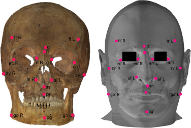
💀 The Skull & Teeth - Cranial Case Crackers
While the pelvis is generally considered the most reliable skeletal region for sex estimation, the skull is a valuable supplementary tool, especially when the pelvis is unavailable or ambiguous. Modern forensic anthropology emphasizes a multifactorial approach, often utilizing a combination of cranial features and other skeletal elements for forensic identification. Combined with dental evidence, the skull provides comprehensive biological profiling capabilities essential for forensic investigations under BSA Section 45 (expert opinion evidence).
📌 Remember: SKULL for major cranial bones - Sphenoid (central), Kull vault (frontal, parietal, occipital), Upper face (maxilla, nasal), Lower jaw (mandible), Lateral temporal bones (22 total bones)
Cranial Sexual Dimorphism
Sexual dimorphism in the skull can achieve 85-90% accuracy when multiple features are systematically assessed using validated methodologies. Male skulls typically display more robust features, larger muscle attachment sites, and greater overall size, while female skulls show more gracile morphology and smoother contours. These assessments require reference to specific, validated methodologies rather than universal fixed values.
- Robust vs. Gracile Features
- Supraorbital ridges: Males prominent, females smooth
- Mastoid processes: Males large (>15mm), females small (<10mm)
- Nuchal crest: Males pronounced, females minimal
- Glabella scoring: 1-5 scale (males 4-5, females 1-2) per Walker methodology
- Mental eminence: Males square, females pointed
- Cranial Capacity Differences
- Male average: 1,450-1,500 cm³
- Female average: 1,300-1,350 cm³
- Overlap zone: 15-20% of population
- Size correlation: r = 0.7 with body size
- Population variation: ±100 cm³ between groups
Loading diagram…
⭐ Clinical Pearl: The combination of 5 cranial features (supraorbital ridge, mastoid process, nuchal crest, mental eminence, and glabella) can achieve high accuracy in sex determination when using validated methodologies, with individual features showing variable accuracy depending on population and assessment method.
Ancestry Assessment from Cranial Morphology
Modern forensic anthropology acknowledges the limitations of traditional ancestry classifications due to increasing population admixture and the continuous nature of human variation. While traditional frameworks can provide general guidance, the emphasis is shifting towards more nuanced, statistical, and population-specific approaches. Ancestry estimation focuses on assessing biological affinity or population substructure rather than assigning discrete categories.
| Ancestry Group | Nasal Aperture | Orbital Shape | Prognathism | Cranial Index | Key Features |
|---|---|---|---|---|---|
| European | Narrow (25-28mm) | Angular | Minimal | 75-80 | High nasal bridge |
| African | Wide (30-35mm) | Rounded | Pronounced | 70-75 | Rectangular orbits |
| Asian | Intermediate (28-32mm) | Rounded | Moderate | 80-85 | Shovel incisors |
| Native American | Intermediate (26-30mm) | Rounded | Minimal | 78-83 | Inca bones |
| Mixed | Variable | Variable | Variable | Variable | Mosaic features |
| %%{init: {'flowchart': {'htmlLabels': true}}}%% | |||||
| flowchart TD |
Ancestry["📋 Ancestry Assessment
• BNS Sec 103• Initial evaluation"]
Affinity["📋 Population Affinity
• Affinity analysis• Morphological grouping"]
Nasal["🔬 Nasal Aperture
• Width measurement• Shape assessment"]
Orbital["🔬 Orbital Morphology
• Eye socket shape• Border analysis"]
Prognathism["🔬 Prognathism
• Jaw projection• Facial profile"]
Stats["🔬 Statistical Analysis
• Data compilation• Probative value"]
Expert["🩺 Expert Opinion
• BSA Sec 45• Forensic conclusion"]
Doc["✅ Documentation
• BNSS Sec 176• Final report"]
Ancestry --> Affinity Affinity --> Nasal Affinity --> Orbital Affinity --> Prognathism Nasal --> Stats Orbital --> Stats Prognathism --> Stats Stats --> Expert Expert --> Doc
style Ancestry fill:#FEF8EC, stroke:#FBECCA, stroke-width:1.5px, rx:12, ry:12, color:#854D0E style Affinity fill:#FEF8EC, stroke:#FBECCA, stroke-width:1.5px, rx:12, ry:12, color:#854D0E style Nasal fill:#FFF7ED, stroke:#FFEED5, stroke-width:1.5px, rx:12, ry:12, color:#C2410C style Orbital fill:#FFF7ED, stroke:#FFEED5, stroke-width:1.5px, rx:12, ry:12, color:#C2410C style Prognathism fill:#FFF7ED, stroke:#FFEED5, stroke-width:1.5px, rx:12, ry:12, color:#C2410C style Stats fill:#FFF7ED, stroke:#FFEED5, stroke-width:1.5px, rx:12, ry:12, color:#C2410C style Expert fill:#F7F5FD, stroke:#F0EDFA, stroke-width:1.5px, rx:12, ry:12, color:#6B21A8 style Doc fill:#F6F5F5, stroke:#E7E6E6, stroke-width:1.5px, rx:12, ry:12, color:#525252
> 💡 **Master This**: Ancestry assessment requires evaluating **multiple cranial features** simultaneously within appropriate population contexts, as no single trait is diagnostic. The accuracy of broad ancestry classification should be contextualized with reference to specific methodologies and populations studied.
### Dental Evidence and Age Estimation
Dental remains provide exceptional preservation and contain multiple age indicators, from development timing in youth to wear patterns in adults. Teeth resist environmental degradation better than bone, often surviving when other skeletal elements are destroyed, making them crucial for **BNS Section 103** (identification of deceased persons).
* **Dental Development Timing**
- First molar eruption: **6 years ±6 months**
- Second molar eruption: **12 years ±1 year**
- Third molar completion: **18-25 years ±2 years**
+ Root development: Continues **2-3 years** post-eruption
+ Population variation: **±6 months** between groups
* **Adult Dental Age Indicators**
- Attrition patterns: **1-2mm** wear per decade
- Secondary dentin formation: Increases with age
- Root translucency: **0.5mm** per decade after **age 20**
+ Accuracy: **±5-8 years** in adults
+ Environmental factors: Diet affects wear rates
> 📌 **Remember**: **TEETH** for dental age assessment - **T**iming of eruption (**±6 months** accuracy), **E**namel formation complete, **E**ruption sequence predictable, **T**hird molars variable (**18-25 years**), **H**istological changes with age
### Cranial Pathology and Individual Characteristics
Pathological conditions and individual variations in cranial morphology provide unique identifying features that can link remains to specific individuals under **BSA Section 9** (relevance of facts forming part of same transaction). These characteristics include congenital anomalies, acquired conditions, and surgical modifications.
* **Congenital Variations**
- Metopic suture persistence: **5-10%** of population
- Wormian bones: **40-60%** frequency in lambdoid suture
- Palatine torus: **20-25%** prevalence, ancestry-dependent
+ Genetic basis: Family clustering patterns
+ Forensic value: Unique individual identifiers
* **Acquired Characteristics**
- Healed fractures: **Callus formation** patterns
- Dental modifications: **Fillings, crowns, extractions**
- Surgical implants: **Plates, screws, prosthetics**
+ Medical records: Direct comparison possible under **BNSS Section 172**
+ Timing assessment: Healing stages indicate chronology
> ⭐ **Clinical Pearl**: Dental records provide the most reliable method for positive identification, with **dental uniqueness** comparable to fingerprints. Even **partial dental remains** can establish identity when antemortem records are available for comparison under **BSA Section 45**.
### Advanced Cranial Analysis Techniques
Modern forensic anthropology employs sophisticated analytical methods for cranial assessment, including geometric morphometrics, 3D imaging, and computer-assisted classification systems that improve accuracy and objectivity for **BNS Section 103** investigations.
* **Geometric Morphometric Analysis**
- **3D landmark coordinates**: **50-100 points** per skull
- Statistical shape analysis: Principal component analysis
- Population databases: **Thousands of specimens**
+ Accuracy improvement: **5-10%** over traditional methods
+ Objectivity: Reduces observer bias
* **Facial Reconstruction Applications**
- Tissue depth standards: **Population-specific measurements**
- Computer modeling: **3D reconstruction software**
- Identification success: **60-70%** recognition rates
+ Investigative tool: Generates leads, not positive ID
+ Media applications: Public assistance in identification under **BNSS Section 41**
```mermaid
%%{init: {'flowchart': {'htmlLabels': true}}}%%
flowchart TD
Start["<b>💀 Advanced Cranial Analysis</b><br><span style='display:block; text-align:left; color:#555'>• BNS Sec 103</span><span style='display:block; text-align:left; color:#555'>• Primary assessment</span>"]
Morph["<b>📐 3D Morphometrics</b><br><span style='display:block; text-align:left; color:#555'>• Structural sizing</span><span style='display:block; text-align:left; color:#555'>• Assessment step</span>"]
Landmark["<b>📍 Landmark Analysis</b><br><span style='display:block; text-align:left; color:#555'>• Coordinate mapping</span><span style='display:block; text-align:left; color:#555'>• Point detection</span>"]
Shape["<b>📊 Statistical Shape</b><br><span style='display:block; text-align:left; color:#555'>• Variance analysis</span><span style='display:block; text-align:left; color:#555'>• Shape comparison</span>"]
Facial["<b>👤 Facial Reconstruction</b><br><span style='display:block; text-align:left; color:#555'>• Identity modeling</span><span style='display:block; text-align:left; color:#555'>• Visual recovery</span>"]
PopDB["<b>📂 Population Database</b><br><span style='display:block; text-align:left; color:#555'>• Regional metrics</span><span style='display:block; text-align:left; color:#555'>• Demographic check</span>"]
Opinion["<b>⚖️ BSA Sec 45 Opinion</b><br><span style='display:block; text-align:left; color:#555'>• Expert testimony</span><span style='display:block; text-align:left; color:#555'>• Forensic findings</span>"]
Doc["<b>📄 BNSS Sec 176 Doc</b><br><span style='display:block; text-align:left; color:#555'>• Evidence records</span><span style='display:block; text-align:left; color:#555'>• Legal compliance</span>"]
Lead["<b>💡 Investigative Lead</b><br><span style='display:block; text-align:left; color:#555'>• Case development</span><span style='display:block; text-align:left; color:#555'>• Identity clues</span>"]
Public["<b>📢 BNSS Sec 41 Public</b><br><span style='display:block; text-align:left; color:#555'>• Public assistance</span><span style='display:block; text-align:left; color:#555'>• Community outreach</span>"]
Start --> Morph
Morph --> Landmark
Morph --> Shape
Morph --> Facial
Landmark --> PopDB
Shape --> PopDB
PopDB --> Opinion
Opinion --> Doc
Facial --> Lead
Lead --> Public
style Start fill:#F7F5FD, stroke:#F0EDFA, stroke-width:1.5px, rx:12, ry:12, color:#6B21A8
style Morph fill:#FEF8EC, stroke:#FBECCA, stroke-width:1.5px, rx:12, ry:12, color:#854D0E
style Landmark fill:#FFF7ED, stroke:#FFEED5, stroke-width:1.5px, rx:12, ry:12, color:#C2410C
style Shape fill:#FFF7ED, stroke:#FFEED5, stroke-width:1.5px, rx:12, ry:12, color:#C2410C
style Facial fill:#FFF7ED, stroke:#FFEED5, stroke-width:1.5px, rx:12, ry:12, color:#C2410C
style PopDB fill:#EEFAFF, stroke:#DAF3FF, stroke-width:1.5px, rx:12, ry:12, color:#0369A1
style Opinion fill:#F1FCF5, stroke:#BEF4D8, stroke-width:1.5px, rx:12, ry:12, color:#166534
style Doc fill:#F6F5F5, stroke:#E7E6E6, stroke-width:1.5px, rx:12, ry:12, color:#525252
style Lead fill:#FDF4F3, stroke:#FCE6E4, stroke-width:1.5px, rx:12, ry:12, color:#B91C1C
style Public fill:#F6F5F5, stroke:#E7E6E6, stroke-width:1.5px, rx:12, ry:12, color:#525252
The skull and teeth together provide comprehensive demographic and individual information available to forensic anthropologists. Understanding cranial morphology, dental development, and pathological variations enables biological profiling and contributes significantly to forensic identification efforts under the BNS 2023 framework.
💀 The Skull & Teeth — Cranial Case Crackers


🦴 Pelvis & Long Bones - Pelvic Pointers & Limb Lengths
The pelvis and long bones represent the most reliable skeletal elements for demographic assessment in forensic anthropology. Pelvic morphology achieves the highest accuracy for sex determination due to functional adaptations for childbirth in females, while long bone measurements provide precise stature estimates through established regression formulae. Understanding these skeletal elements enables accurate biological profiling essential for forensic identification under BNS Section 63 (causing death by negligence) and BNSS Section 174 (police investigation procedures).
📌 Remember: PELVIS for sex determination features - Pubic angle (>90° female, <90° male), Examination of auricular surface, Large sciatic notch (female wider/U-shaped), Ventral arc present (female), Ischiopubic ramus medial aspect (thin in female), Subpubic contour (concave in female)
Pelvic Sexual Dimorphism
The pelvis demonstrates the most pronounced sexual dimorphism in the human skeleton, reflecting functional adaptations for parturition in females. Multiple morphological features can be assessed independently, with combined analysis achieving 80-90% accuracy in well-preserved specimens for sex determination.
- Primary Sex Indicators
- Subpubic angle: Males <90°, females >90°
- Greater sciatic notch: Males narrow/V-shaped, females wide/U-shaped
- Ventral arc: Present in 96% of females, absent in males
- Subpubic contour: Concave in 92% of females, 7% of males
- Ischiopubic ramus medial aspect: Thin/sharp in females, thick/blunt in males
- Secondary Sex Indicators
- Pelvic inlet shape: Males heart-shaped, females oval
- Acetabulum diameter: Males >52mm, females <50mm (population-specific)
- Ischiopubic ramus: Males thick/robust, females thin/gracile
- Sacral curvature: Females more pronounced (variable)
- Iliac blade: Females more flared laterally (subjective assessment)
⭐ Clinical Pearl: The Phenice method using three pelvic features (ventral arc, subpubic contour, ischiopubic ramus medial aspect) achieves 96% accuracy in sex determination when well-preserved, though accuracy varies with population groups and preservation conditions.
Long Bone Stature Estimation
Long bone measurements provide the most accurate method for stature estimation from skeletal remains, utilizing regression formulae developed from documented skeletal collections. Accuracy varies by bone used and population ancestry, with femur measurements providing optimal results.
| Bone | Measurement | Accuracy (±cm) | Population Specificity | Regression Formula Example |
|---|---|---|---|---|
| Femur | Maximum length | ±3.27 | High | 2.38 × femur + 61.41 |
| Tibia | Maximum length | ±3.57 | High | 2.52 × tibia + 78.62 |
| Humerus | Maximum length | ±4.05 | Moderate | 2.89 × humerus + 78.10 |
| Radius | Maximum length | ±4.24 | Moderate | 3.79 × radius + 79.42 |
| Ulna | Maximum length | ±4.30 | Moderate | 3.76 × ulna + 75.55 |
| %%{init: {'flowchart': {'htmlLabels': true}}}%% | ||||
| flowchart TD |
Start["🦴 Skeletal Remains
• Discovery phase• Remains found"]
Investigate["📋 BNS Sec 174
• Police inquiry• Initial process"]
Exam["🔬 Forensic Exam
• Anthropology work• Lab analysis"]
SexDet["🧬 Pelvic Analysis
• Sex determination• Os coxae study"]
Stature["📏 Long Bone
• Stature profile• Bone measurement"]
Phenice["📋 Phenice Method
• Morphological check• Assessment tool"]
Formula["🧮 Population Math
• Specific formulae• Height calculation"]
Opinion["⚖️ BSA Sec 45
• Expert opinion• Formal report"]
Testimony["🏛️ BNSS Sec 293
• Court testimony• Legal evidence"]
Start --> Investigate Investigate --> Exam Exam --> SexDet Exam --> Stature SexDet --> Phenice Stature --> Formula Phenice --> Opinion Formula --> Opinion Opinion --> Testimony
style Start fill:#F7F5FD, stroke:#F0EDFA, stroke-width:1.5px, rx:12, ry:12, color:#6B21A8 style Investigate fill:#FEF8EC, stroke:#FBECCA, stroke-width:1.5px, rx:12, ry:12, color:#854D0E style Exam fill:#FFF7ED, stroke:#FFEED5, stroke-width:1.5px, rx:12, ry:12, color:#C2410C style SexDet fill:#FFF7ED, stroke:#FFEED5, stroke-width:1.5px, rx:12, ry:12, color:#C2410C style Stature fill:#FFF7ED, stroke:#FFEED5, stroke-width:1.5px, rx:12, ry:12, color:#C2410C style Phenice fill:#FEF8EC, stroke:#FBECCA, stroke-width:1.5px, rx:12, ry:12, color:#854D0E style Formula fill:#FFF7ED, stroke:#FFEED5, stroke-width:1.5px, rx:12, ry:12, color:#C2410C style Opinion fill:#F6F5F5, stroke:#E7E6E6, stroke-width:1.5px, rx:12, ry:12, color:#525252 style Testimony fill:#FDF4F3, stroke:#FCE6E4, stroke-width:1.5px, rx:12, ry:12, color:#B91C1C
> 💡 **Master This**: Femur maximum length provides the most accurate stature estimates, with **Trotter-Gleser formulae** achieving **±3.27cm** accuracy. When multiple bones are available, using **femur + tibia** measurements together improves precision to **±2.8cm**.

### Population-Specific Considerations

Stature estimation accuracy depends significantly on population ancestry, as body proportions vary between groups. Modern forensic practice requires population-specific formulae or adjustment factors to achieve optimal accuracy.

* **Population Variation in Body Proportions**
  - African ancestry: Longer limbs relative to trunk (**+2-3cm** adjustment)
  - Asian ancestry: Shorter limbs relative to trunk (**-1-2cm** adjustment)
  - European ancestry: Intermediate proportions (baseline formulae)
    + Secular trends: **+1-2cm** per generation in developed populations
    + Sex differences: **10-15cm** average height difference
* **Modern Formulae Applications**
  - Fordisc software: Population-specific calculations
  - ADBOU standards: Contemporary American populations
  - International databases: Global population coverage
    + Accuracy improvement: **15-20%** over generic formulae
    + Database size: **>10,000** documented cases


> 📌 **Remember**: **STATURE** for estimation principles - **S**ex-specific formulae required, **T**rotter-Gleser most common, **A**ncestry affects accuracy, **T**ibia second choice after femur, **U**se multiple bones when possible, **R**egression error ±**3-4cm**, **E**nvironmental factors influence growth

### Pathological Conditions and Trauma

Long bones and pelvis frequently preserve evidence of pathological conditions, trauma, and activity patterns that provide additional information for forensic identification and cause of death determination under **BNS Section 101** (culpable homicide) investigations.

* **Degenerative Changes**
  - Osteoarthritis: Joint surface **eburnation** and **osteophyte** formation
  - Vertebral compression: **Height loss** and **wedging**
  - Hip dysplasia: **Acetabular** modifications and **femoral head** changes
    + Age correlation: Severity increases with advancing age
    + Activity patterns: Occupational stress markers
* **Trauma Indicators**
  - Fracture patterns: **Antemortem** vs. **perimortem** vs. **postmortem**
  - Healing stages: **Callus formation** timing (**2-6 weeks**)
  - Surgical interventions: **Hardware placement** and **bone modifications**
    + Timing assessment: Healing progression indicates chronology
    + Cause determination: Force direction and magnitude

> ⭐ **Clinical Pearl**: Pelvic fractures in forensic contexts often indicate **high-energy trauma** such as motor vehicle accidents or falls from height, with **bilateral fractures** suggesting **>50% mortality** in clinical settings due to associated injuries.

### Advanced Analytical Techniques

Modern forensic anthropology employs sophisticated methods for pelvic and long bone analysis, including 3D imaging, geometric morphometrics, and machine learning algorithms that improve accuracy and reduce observer error in **BSA Section 45** expert testimony.

* **3D Imaging Applications**
  - CT scanning: **Sub-millimeter** resolution measurements
  - Photogrammetry: **Field documentation** and **virtual analysis**
  - Surface scanning: **Morphological analysis** without handling
    + Measurement precision: **±0.1mm** accuracy possible
    + Database integration: Digital specimen comparison
* **Machine Learning Classification**
  - Neural networks: **Pattern recognition** for sex determination
  - Support vector machines: **Multi-feature** classification
  - Random forests: **Ancestry assessment** algorithms
    + Accuracy improvement: **2-5%** over traditional methods
    + Objectivity: Reduced observer bias and error


The pelvis and long bones provide the foundation for accurate demographic assessment in forensic anthropology. Understanding sexual dimorphism, population variation, and measurement techniques enables precise biological profiling that contributes significantly to forensic identification and medicolegal investigations under the **BNS 2023** framework.
🦴 Pelvis & Long Bones — Pelvic Pointers & Limb Lengths


⚡ High‑Yield Points - Biggest Takeaways
Essential Accuracy Benchmarks
Understanding accuracy limitations and optimal methods for each demographic parameter enables appropriate confidence levels in forensic reporting and testimony under BSA Section 45 expert evidence requirements. These benchmarks guide method selection and interpretation of results in medicolegal contexts.
⭐ Clinical Pearl: Sex determination from pelvis achieves 95-98% accuracy using multiple features, while skull analysis reaches 85-90% accuracy. Combined pelvic and cranial assessment approaches 99% reliability in adult remains. However, modern forensic anthropology heavily relies on DNA analysis for positive identification and to provide highly accurate information regarding sex and ancestry, significantly impacting practice standards.
- Demographic Assessment Accuracy Hierarchy
- Sex determination: Pelvis (95-98%) > Skull (85-90%) > Long bones (80-85%)
- Age estimation: Subadults (±1-2 years) > Young adults (±3-5 years) > Older adults (±8-12 years)
- Stature estimation: Femur (±3.27cm) > Tibia (±3.57cm) > Humerus (±4.05cm)
- Ancestry assessment: 75-80% accuracy with multiple cranial features
💡 Master This: Age estimation accuracy decreases dramatically after age 30, requiring multiple methods and wider confidence intervals. Modern forensic anthropology emphasizes population-specific standards and reporting ages as ranges with appropriate confidence intervals, not point estimates, with error margins increasing ±2-3 years per decade of life.
Critical Timing Sequences
Memorizing key developmental and degenerative timing enables rapid age assessment and appropriate method selection based on available skeletal elements, with findings admissible under BSA Section 45.
📌 Remember: FUSION sequence for epiphyseal closure represents general guidelines with individual variation - Fibula head (16-18), Ulnar olecranon (14-16), Sacral segments (18-25+), Iliac crest (20-23+), Occipital basilar (18-20), Nasal bones (birth). These are simplified representations requiring multiple indicators for adult age estimation.
| Age Range | Primary Method | Key Landmarks | Accuracy | Population Variation |
|---|---|---|---|---|
| Fetal | Bone length | Femur 7-35mm | ±2 weeks | Minimal |
| 0-6 years | Dental eruption | First molars 6 years | ±6 months | ±3 months |
| 6-18 years | Epiphyseal fusion | Multiple sites | ±1-2 years | ±6 months |
| 18-30 years | Late fusion | Clavicle 25-30 | ±3-5 years | ±2 years |
| 30+ years | Degenerative changes | Multiple indicators | ±8-12 years | ±5 years |
Understanding population variation prevents misclassification and improves accuracy in diverse forensic contexts. Modern populations show increasing admixture, requiring nuanced approaches to ancestry assessment under BNSS investigation protocols.
Loading diagram…
⭐ Clinical Pearl: Ancestry assessment requires multiple features due to population admixture. Modern forensic anthropology acknowledges significant impact of population admixture - use probabilistic approach with multiple features rather than definitive single traits for 75-80% accuracy in broad classification.
- Key Population Markers (with admixture considerations)
- Asian ancestry: Shovel-shaped incisors (historically 85% frequency), rounded orbits
- African ancestry: Rectangular orbits, pronounced prognathism, wide nasal aperture (historically >30mm)
- European ancestry: Narrow nasal aperture (historically <28mm), minimal prognathism, angular orbits
- Modern admixture: 20-40% of individuals show mixed features requiring complex assessment
- Geographic variation: Significant within-group diversity challenges strict measurements
Taphonomic Considerations
Understanding postmortem changes enables accurate interpretation of skeletal modifications and prevents misidentification of trauma or pathology in BNS Section 174 death investigations.
📌 Remember: CHANGES in bone after death - Cracking from drying, Heating discoloration, Animal modification, Natural weathering, Groundwater staining, Erosion patterns, Soil chemistry effects
- Postmortem Interval Indicators
- Fresh bone: Greasy texture, intact periosteum
- Dry bone: Chalky appearance, surface cracking
- Weathered bone: Bleaching, surface exfoliation
- Timeline factors: Climate, burial environment, body size
- Preservation variation: Months to decades depending on conditions
Legal and Ethical Framework
Forensic anthropology operates within strict legal and ethical guidelines under the 2024 legal framework that govern evidence handling, testimony standards, and professional responsibilities.
⭐ Clinical Pearl: Chain of custody documentation is critical for BSA Section 63 admissibility requirements. Every handling, analysis, and storage event must be recorded with date, time, personnel, and purpose to maintain evidence integrity under BNSS procedures.
- Professional Standards
- ABFA certification: Board-certified forensic anthropologists
- Laboratory accreditation: ISO/IEC 17025 standards
- Continuing education: Annual requirements for certification maintenance
- Testimony qualifications: BSA Section 45 expert witness standards
- Report writing: Scientific methodology and statistical interpretation
Technology Integration
Modern forensic anthropology increasingly relies on advanced technology for analysis, documentation, and database comparison, improving accuracy and efficiency while meeting BSA Section 45 scientific evidence standards.
- Digital Documentation
- 3D scanning: Sub-millimeter precision measurements
- Photogrammetry: Field documentation and virtual analysis
- Database comparison: Fordisc, ADBOU, international collections
- Measurement standardization: Reduced observer error
- Remote consultation: Expert collaboration across distances
💡 Master This: Technology enhances but never replaces fundamental anatomical knowledge. Understanding skeletal biology, population variation, and taphonomic processes remains essential for accurate interpretation of digital analysis results, especially when DNA analysis provides definitive identification and demographic information.
These high-yield concepts form the essential knowledge base for forensic anthropological practice under the 2024 legal framework, enabling accurate biological profiling that serves BNS and BNSS justice system needs while maintaining BSA scientific rigor and professional standards, with emphasis on population-specific standards, confidence intervals, and DNA integration for modern practice.
⚡ High‑Yield Points — Biggest Takeaways


🔬 Advanced Integration - The Connectome Architecture
📌 Remember: INTEGRATE for comprehensive analysis - Individual variation assessment, New technology applications, Taphonomic factor evaluation, Environmental context analysis, Genetic ancestry considerations, Recovery context documentation, Age-related changes, Trauma pattern interpretation, Evidence correlation
Multi-System Skeletal Analysis
Comprehensive skeletal analysis requires understanding interactions between different anatomical systems and how pathological conditions, trauma, and environmental factors affect multiple skeletal elements simultaneously.
- Systemic Disease Manifestations
- Osteoporosis: Vertebral compression (>20% height loss), femoral neck thinning
- Osteoarthritis: Joint space narrowing, osteophyte formation, subchondral sclerosis
- Metabolic disorders: Bone density changes, growth disruption, mineralization defects
- Multi-element involvement: Spine, pelvis, long bones affected differently
- Age-related progression: Severity correlates with chronological age
- Trauma Pattern Recognition
- Blunt force: Radiating fractures, butterfly fragments, plastic deformation
- Video documentation and biomechanical analysis of falls from height can provide valuable evidence, revealing distinct patterns of impact on different ground surfaces
- Sharp force: Clean margins, kerf marks, striation patterns
- Research has shown that the analysis of striation patterns on bone can aid in the identification of weapon types and the materials used for maintenance or sharpening. This can offer additional evidentiary evidence in forensic investigations
- Studies have shown that cut marks on bone, even after heat exposure, remain recognizable and do not alter considerably, allowing for continued analysis in forensic casework
- Ballistic trauma: Entrance/exit characteristics, lead deposition, fracture patterns
- Force direction: Fracture morphology indicates impact angle
- Timing assessment: Healing stages distinguish antemortem/perimortem
- Blunt force: Radiating fractures, butterfly fragments, plastic deformation
Loading diagram…
⭐ Clinical Pearl: Bilateral symmetry assessment reveals systemic conditions versus localized trauma. Osteoarthritis typically shows bilateral involvement with asymmetric severity, while acute trauma demonstrates unilateral patterns with specific force characteristics.
Technology-Enhanced Analysis
Modern forensic anthropology integrates multiple technological approaches to improve accuracy, reduce analysis time, and enable remote collaboration between experts worldwide.
| Technology | Application | Clinical Benefits | Cost Factor | Training Required |
|---|---|---|---|---|
| 3D CT Scanning | Internal structure analysis | Reveals osteotomy edges, bone graft integration, early hardware failure | High | Moderate |
| Photogrammetry | Field documentation | 10-15% | Low | Minimal |
| Geometric Morphometrics | Shape analysis | 5-10% | Moderate | High |
| Isotope Analysis | Geographic origin | Variable | High | High |
| Ancient DNA | Individual identification | 99%+ | Very High | Specialized |
- Fordisc 3.0: >4,000 documented specimens for comparison
- ADBOU standards: Contemporary American population data
- International databases: Global population coverage expanding
- Statistical validation: Cross-validation with known samples
- Population specificity: Ancestry-specific algorithms improve accuracy
💡 Master This: Technology amplifies expertise but cannot replace fundamental knowledge. CT scans are particularly effective at revealing osteotomy edges, bone graft integration, and early hardware failure, which may not be apparent on conventional radiographs. However, CT scans may sometimes over-diagnose non-union due to differing definitions of union. Additionally, while the radiation dose for peripheral limb CT is relatively low, it increases significantly for areas closer to radiosensitive tissue, such as the shoulder and hip, which requires careful consideration during imaging.
Environmental Context Integration
Understanding environmental factors affecting skeletal preservation, modification, and recovery enables accurate interpretation of postmortem changes and optimal evidence collection strategies.
- Preservation Environment Effects
- Acidic soils: Bone dissolution, mineral loss, fragmentation
- Alkaline conditions: Enhanced preservation, mineral replacement
- Waterlogged environments: Soft tissue preservation, bone softening
- Temperature effects: Freeze-thaw cycles cause mechanical damage
- Humidity variation: Cracking patterns from desiccation
- Recovery Context Documentation
- GPS coordinates: Sub-meter accuracy for scene mapping
- Photographic documentation: Multiple angles, scale references
- Stratigraphic relationships: Temporal sequence reconstruction
- Associated artifacts: Cultural materials provide chronological context
- Botanical evidence: Seasonal indicators and time since deposition
📌 Remember: CONTEXT for environmental factors - Climate effects on preservation, Oxygen availability impacts decomposition, Natural processes modify remains, Temperature affects timing, Erosion patterns indicate exposure, Xenobiotic contamination, Taphonomic signatures
Interdisciplinary Collaboration Framework
Effective forensic anthropology requires seamless integration with multiple disciplines, each contributing specialized expertise to comprehensive case resolution under the BSA Sec 45 expert evidence framework.
- Core Collaboration Partners
- Forensic pathologists: Cause of death determination, soft tissue analysis
- Forensic odontologists: Dental identification, bite mark analysis
- DNA analysts: Genetic identification, kinship analysis
- Crime scene investigators: Context documentation, evidence recovery
- Forensic entomologists: Postmortem interval estimation
- Communication Protocols
- Standardized terminology: Consistent reporting across disciplines
- Evidence sharing: Chain of custody maintenance per BNSS Sec 61
- Joint consultations: Complex case collaboration
- Quality assurance: Peer review processes
- Continuing education: Interdisciplinary training programs
⭐ Clinical Pearl: Collaborative analysis improves case resolution rates by 25-30% compared to single-discipline approaches. Early consultation between specialists prevents evidence loss and analytical conflicts that can compromise investigations under BNS framework requirements.
Cutting-Edge Research Applications
Emerging technologies and methodologies continue expanding forensic anthropology capabilities, offering new approaches to challenging identification problems and cold cases within the BSA evidence standards.
- Isotope Analysis Applications
- Strontium isotopes: Geographic origin determination (±100km resolution)
- Oxygen isotopes: Climate zone identification
- Carbon/nitrogen isotopes: Dietary reconstruction
- Childhood mobility: Tooth enamel preserves early life signatures
- Adult residence: Bone collagen reflects recent years
- Facial Reconstruction Technology
- Computer modeling: 3D soft tissue prediction
- Population-specific standards: Tissue depth databases
- Recognition success: 60-70% identification rates
- Investigative tool: Generates leads, not positive identification
- Media applications: Public assistance in cold cases
Loading diagram…
💡 Master This: Emerging technologies expand identification possibilities but require rigorous validation before forensic application under BSA standards. Isotope analysis shows promise for geographic origin determination, while facial reconstruction serves as investigative tool rather than identification method. MRI and nuclear imaging may be useful for detecting soft tissue complications, though their role in non-union prediction remains limited.
Advanced integration in forensic anthropology represents the evolution from descriptive analysis to predictive science, where understanding complex interactions between biological, environmental, and technological factors enables comprehensive case resolution that serves both scientific advancement and justice system needs under the BNS, BNSS, and BSA framework.
🔬 Advanced Integration — The Connectome Architecture


🎯 Mastery Framework - Rapid Reference Arsenal
Essential Quick-Reference Matrix
Master these core values for immediate field and laboratory application, enabling rapid demographic assessment without extensive calculation or reference consultation.
⭐ Clinical Pearl: While the 95-85-75 Rule provides general guidelines for pelvis sex determination (95% accuracy), skull sex determination (85% accuracy), and ancestry assessment (75% accuracy), these are broad estimates. Actual accuracy varies significantly based on population, methodology, and specific features analyzed. Modern forensic anthropology emphasizes statistical methods and population-specific data for more precise estimations.
| Assessment | Primary Method | Accuracy | Key Threshold | Population Variation |
|---|---|---|---|---|
| Sex - Pelvis | Subpubic angle | 95-98% | Range of angles | ±5° |
| Sex - Skull | Multiple features | 85-90% | 5-feature combo | ±10% |
| Age - Subadult | Dental development | ±1 year | 6-year molars | ±6 months |
| Age - Adult | Multifactorial approach | ±8-12 years | 30+ years | ±5 years |
| Stature | Population-specific formulas | ±3.27cm | Population-dependent | ±2cm ancestry |
| Ancestry | Multiple skeletal traits | 75-80% | Statistical analysis | High admixture |
Critical Measurement Standards
The Standards for Data Collection from Human Skeletal Remains by Buikstra and Ubelaker (1994) provides foundational guidelines, though ongoing research continues to refine these standards. A 2020 study highlighted that 8 out of 15 measurements differed significantly when modified definitions were employed, emphasizing that data collected using different definitions should not be used interchangeably.
- Cranial Measurements
- Maximum cranial length: Glabella to opisthocranion
- Maximum cranial breadth: Euryon to euryon
- Nasal aperture width: Widest point of piriform aperture
- Cranial index: (Breadth/Length) × 100
- Nasal index: (Width/Height) × 100
- Postcranial Measurements
- Femur maximum length: Femoral head to medial condyle
- Humerus maximum length: Humeral head to trochlea
- Pelvic measurements: Internationally recognized landmarks for sex assessment
- Measurement precision: ±1mm required for accuracy
- Instrument calibration: Regular verification essential
Loading diagram…
💡 Master This: Measurement consistency is critical for database comparison. Use internationally recognized landmarks, calibrated instruments, and multiple measurements averaged for optimal accuracy. While inter-observer error <2% is aspirational, rigorous training and statistical methods help minimize both intra- and inter-observer error.
Trauma Recognition Patterns
Rapid trauma assessment enables immediate cause-of-death insights and guides further investigation priorities under BNS homicide provisions.
- Blunt Force Trauma Signatures
- Radiating fractures: Star pattern from impact point
- Concentric fractures: Circular pattern around impact
- Plastic deformation: Bending without complete fracture
- Timing indicators: Sharp edges = perimortem, rounded edges = antemortem
- Force estimation: Fracture pattern complexity correlates with impact energy
- Sharp Force Trauma Characteristics
- Kerf marks: Tool width and blade characteristics
- False start marks: Hesitation or repositioning
- Bone chips: Direction indicates cutting angle
- Tool identification: Microscopic analysis of cut surfaces
- Sequence reconstruction: Overlapping cuts show temporal order
⭐ Clinical Pearl: Perimortem trauma shows sharp fracture edges with no healing, while antemortem trauma demonstrates callus formation and rounded edges. Postmortem damage appears different colored and brittle.
Population-Specific Quick Guides
Ancestry assessment utilizes multiple skeletal traits, dental and postcranial features, employing sophisticated statistical methods and population databases. Ancestry relates to biological geographic origin and genetic heritage, distinct from socially constructed concepts.
📌 Remember: ANCESTRY markers - Asian shovel incisors (85%), Narrow nasal aperture Europeans (<28mm), Caribbean/African wide aperture (>30mm), European angular orbits, Square mental eminence males, Tibial platycnemia varies, Rounded orbits Asian/African, Y-shaped sutures variable
- Cranial Feature Frequencies
- Shovel-shaped incisors: Asian (85%), European (15%), African (10%)
- Carabelli's cusp: European (60%), Asian (40%), African (30%)
- Three-rooted molars: Asian (20%), African (3%), European (<1%)
- Nasal spine: European prominent, Asian intermediate, African minimal
- Prognathism: African pronounced, Asian moderate, European minimal
Legal Testimony Framework
Court testimony under BSA evidence standards requires clear communication of scientific findings with appropriate confidence levels and limitations, following BNSS procedural requirements.
- Testimony Structure
- Qualifications: Education, experience, certifications
- Methodology: Scientific basis for conclusions under BSA standards
- Findings: Specific measurements and statistical probabilities
- Limitations: Confidence intervals and population variation
- Alternative explanations: Differential diagnosis considerations
- Communication Guidelines
- Avoid absolute statements: Use "consistent with" rather than "proves"
- Quantify uncertainty: Provide statistical confidence levels
- Explain methodology: Peer-reviewed scientific basis per BSA requirements
- Visual aids: Photographs, diagrams, comparison materials
- Professional demeanor: Objective, factual, non-advocacy
⭐ Clinical Pearl: Effective testimony under BSA standards combines scientific accuracy with clear communication. Use percentage probabilities rather than absolute statements, and always acknowledge limitations and alternative interpretations to maintain credibility.
Quality Assurance Protocols
Maintaining analytical accuracy requires systematic quality control measures and continuous professional development, ensuring compliance with BSA evidence standards and BNSS procedural requirements.
- Laboratory Standards
- Measurement verification: 10% re-measurement protocol
- Peer review: Complex cases require second opinion
- Documentation: Complete chain of custody per BNSS requirements
- Equipment calibration: Monthly verification of instruments
- Proficiency testing: Annual blind sample analysis
- Professional Development
- Continuing education: 40 hours annually for certification
- Conference participation: Current research integration
- Method validation: New technique evaluation before implementation
- Error rate monitoring: Track accuracy across case types
- Feedback integration: Learn from case outcomes and peer review
Loading diagram…
This mastery framework provides the essential tools for confident, accurate forensic anthropological practice under the 2024 legal framework, enabling rapid assessment while maintaining scientific rigor and professional standards required for medicolegal applications under BNS, BNSS, and BSA provisions.
🎯 Mastery Framework — Rapid Reference Arsenal

Unlock the full lesson and continue reading
Signup to continue reading this lesson and unlimited access questions, flashcards, AI notes, and more


Have doubts about this lesson?
Ask Rezzy, your AI Study Partner, to explain anything you didn't understand
Everything you need for NEET-PG prep
Get full Oncourse access with lessons, practice questions, flashcards and AI study tools.
Scan to download app