
Contrast-Enhanced Ultrasound — Flashcards

Tissue harmonic imaging uses higher harmonic frequencies generated by propagation of the _____ beam through tissue
Which stages of CE based on USG imaging shows univesicular fluid collection?_____
Optison and Levovist are _____ contrast agents
In FAST, Subxiphoid transverse view: assess for pericardial effusion and _____ injuries
Which stage of Cystic Echinococcosis (CE) on ultrasound shows a calcified wall?
Which mode of USG is preferred in echocardiography?_____
_____ sign is seen in normal lung on M-mode ultrasound
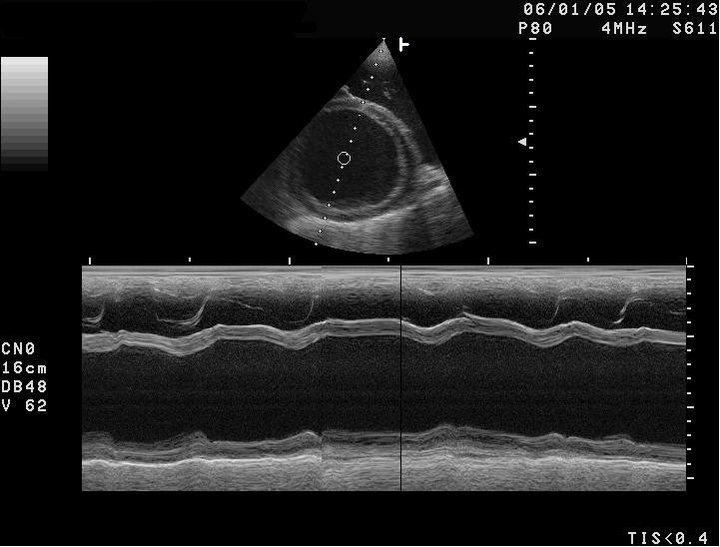
In the following M-mode USG, the X-axis represents _____ and the echo intensity is represented as brightness.
_____ sign is seen in pneumothorax on M-mode ultrasound
_____ sign is seen in chronic cholecystitis with cholelithiasis
Contrast-Enhanced Ultrasound Flashcards for NEET-PG
Study 10 flashcards on Contrast-Enhanced Ultrasound for NEET-PG Radiology. These active recall cards cover the key concepts, clinical associations, and high-yield facts from this chapter of Ultrasound. Each card is designed to test your understanding rather than just recognition, building stronger and more durable memories for exam day.
For personalised spaced repetition scheduling and unlimited flashcards, download the Oncourse app.
Frequently Asked Questions
Are Contrast-Enhanced Ultrasound flashcards free?
How many flashcards does this chapter have?
How should I use these flashcards for NEET-PG?
Are there more flashcards for Ultrasound?
Want unlimited flashcards?
Get full access to all flashcards, spaced repetition, and progress tracking.
Scan to download app