
Cross-sectional Anatomy: Neck — Flashcards

Bulge in posterior pharyngeal wall on x-ray is seen _____laterally in acute retropharyngeal abscess
Which x-ray view shows all paranasal air sinuses?_____
_____ sinuses are seen best with Caldwell's view
Widening of the pre_____ soft tissue shadow, and loss of cervical lordosis is seen in x-ray of retropharyngeal abscess
The radiological hilum is formed by what 3 structures?_____
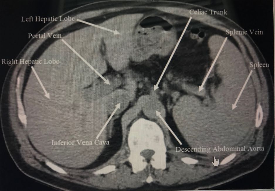
What vertebral level is this CT image taken at? _____
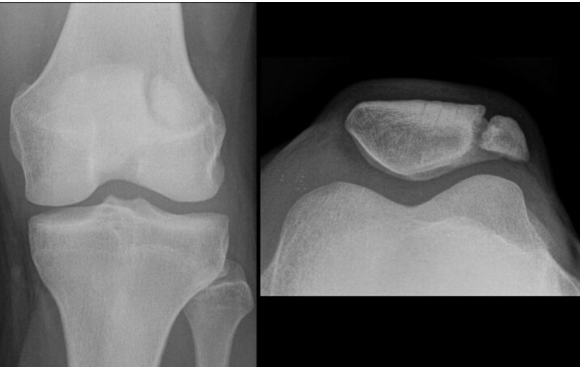
The given X-Ray shows a _____ patella
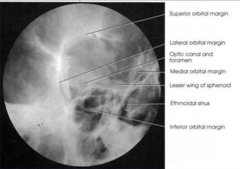
The _____ view in an X-ray scan helps in the visualization of the optic foramina.
72af9e99fc56410ab3d35293d5de0c5f-ao-3
412afda228c940ad8b9196bbffd651cb-ao-1
Cross-sectional Anatomy: Neck Flashcards for NEET-PG
Study 10 flashcards on Cross-sectional Anatomy: Neck for NEET-PG Radiology. These active recall cards cover the key concepts, clinical associations, and high-yield facts from this chapter of Radiological Anatomy. Each card is designed to test your understanding rather than just recognition, building stronger and more durable memories for exam day.
For personalised spaced repetition scheduling and unlimited flashcards, download the Oncourse app.
Frequently Asked Questions
Are Cross-sectional Anatomy: Neck flashcards free?
How many flashcards does this chapter have?
How should I use these flashcards for NEET-PG?
Are there more flashcards for Radiological Anatomy?
Want unlimited flashcards?
Get full access to all flashcards, spaced repetition, and progress tracking.
Scan to download app