
Cross-sectional Anatomy: Abdomen and Pelvis — Flashcards

A _____ bladder is one whose normal round or ovoid shape has been extrinsically compressed into a narrow, vertical orientation (e.g., by pelvic lipomatosis).
_____ sign is the anterior interposition of the colon to the liver reaching the under-surface of the right hemidiaphragm
The key area seen in _____ view is the antrum and upper part of the attic.
5db51bc822474d7784e09837a0848bcf-ao-2
Radiological features of scurvy: _____: Lucent metaphyseal band underlying frankel line. Also called Scorbutic zone
Salt and pepper appearance of the skull is a radiological feature of _____
Radiographic view of sinuses include: 1. _____ view: Maxillary sinuses2. Caldwell's view: Frontal sinuses3. Submentovertical view: Sphenoid, ethmoid and posterior sinus, seen best in that order.4. Right and left oblique views 5. Lateral view
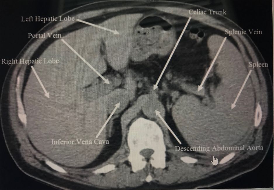
What vertebral level is this CT image taken at? _____
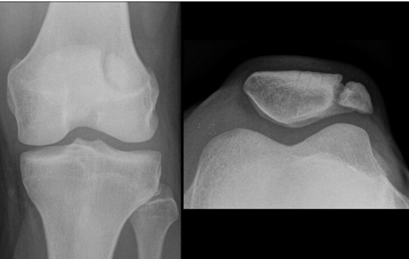
The given X-Ray shows a _____ patella
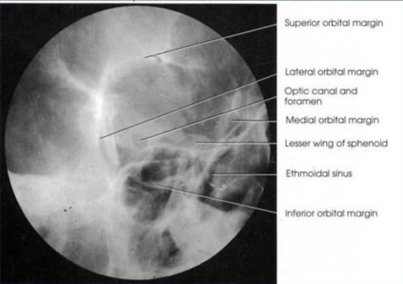
The _____ view in an X-ray scan helps in the visualization of the optic foramina.
Cross-sectional Anatomy: Abdomen and Pelvis Flashcards for NEET-PG
Study 10 flashcards on Cross-sectional Anatomy: Abdomen and Pelvis for NEET-PG Radiology. These active recall cards cover the key concepts, clinical associations, and high-yield facts from this chapter of Radiological Anatomy. Each card is designed to test your understanding rather than just recognition, building stronger and more durable memories for exam day.
For personalised spaced repetition scheduling and unlimited flashcards, download the Oncourse app.
Frequently Asked Questions
Are Cross-sectional Anatomy: Abdomen and Pelvis flashcards free?
How many flashcards does this chapter have?
How should I use these flashcards for NEET-PG?
Are there more flashcards for Radiological Anatomy?
Want unlimited flashcards?
Get full access to all flashcards, spaced repetition, and progress tracking.
Scan to download app