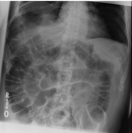
_____ seen in X ray is a focal area of adynamic ileus close to an intra-abdominal inflammatory process
#2
Whirl sign on abdominal CT is diagnostic of _____
#3
_____ sign is seen in CT because in acute pancreatitis perinephric fat is not involved
#4
_____ volvulus is associated with the "coffee bean" sign on X-ray
#5
In _____ sign or double wall sign, inner mucosal and outer serosal layers of bowel are enhanced in pneumoperitoneum
#6
_____ is the initial modality of choice for diagnosing a case of suspected chronic pancreatitis.
#7
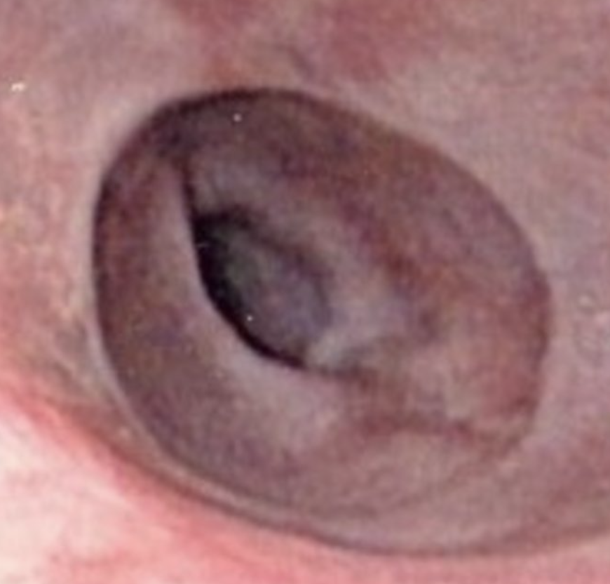
The given image shows the presence of a _____ in lower end of esophagus.
#8
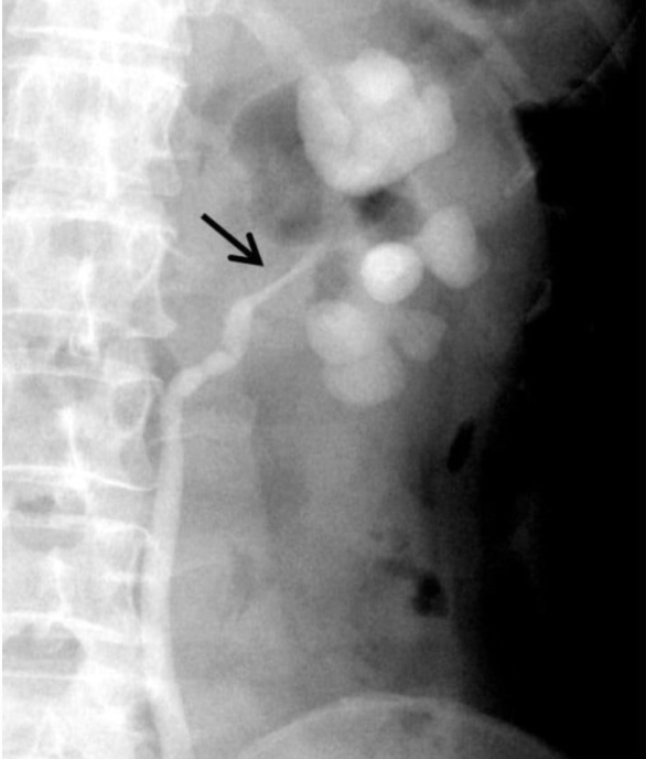
The given Retrograde ureterogram showing dilated clubbed calyces with stricture of the left ureteropelvic junction is suggestive of _____
#9
In _____ sign or double wall sign, inner mucosal and outer serosal layers of bowel are enhanced in pneumoperitoneum
#10
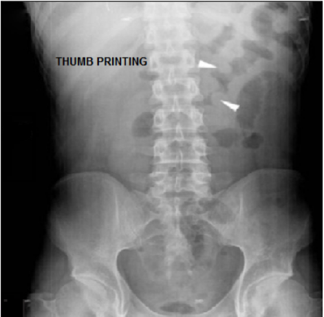
Thumbprinting sign is suggestive of _____
Acute Abdomen Imaging Indian Medical PG Flashcards - Medical Study Cards
Master Acute Abdomen Imaging with OnCourse flashcards. These spaced repetition flashcards are designed for medical students preparing for NEET PG, USMLE Step 1, USMLE Step 2, MBBS exams, and other medical licensing examinations.
Acute Abdomen Imaging Flashcard Deck - 10 Cards
Flashcard 1: _____ seen in X ray is a focal area of adynamic ileus close to an intra-abdominal inflammatory process
Answer: Sentinel loop
Flashcard 2: Whirl sign on abdominal CT is diagnostic of _____
Answer: sigmoid volvulus
Flashcard 3: _____ sign is seen in CT because in acute pancreatitis perinephric fat is not involved
Answer: Preserved renal halo
Flashcard 4: _____ volvulus is associated with the "coffee bean" sign on X-ray
Answer: Sigmoid
Flashcard 5: In _____ sign or double wall sign, inner mucosal and outer serosal layers of bowel are enhanced in pneumoperitoneum
Answer: Rigler's
Flashcard 6: _____ is the initial modality of choice for diagnosing a case of suspected chronic pancreatitis.
Answer: Abdominal CT scan
Flashcard 7: The given image shows the presence of a _____ in lower end of esophagus.
Answer: Schatzki’s ring
Flashcard 8: The given Retrograde ureterogram showing dilated clubbed calyces with stricture of the left ureteropelvic junction is suggestive of _____
Answer: Hydronephrosis
Flashcard 9: In _____ sign or double wall sign, inner mucosal and outer serosal layers of bowel are enhanced in pneumoperitoneum
Answer: Rigler's
Flashcard 10: Thumbprinting sign is suggestive of _____