
Inflammatory Dermatoses — Flashcards

Epidermis of an orf lesion shows ballooning degeneration of keratinocytes with eosinophilic _____ inclusions
_____ nevus is a non-nested dermal infiltration of highly dendritic heavily pigmented nevus cells
In **Staphylococcal Scalded-Skin Syndrome** (SSSS), the exfoliative toxin destroys _____ in the stratum granulosum.
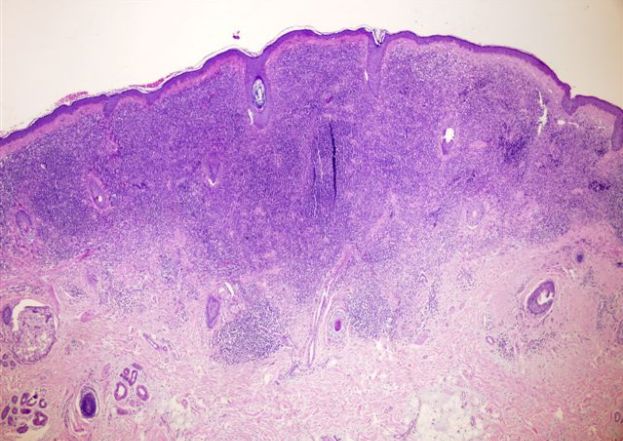
Lichen planus is characterized histologically by inflammation of the _____ junction with a 'saw-tooth' appearance
_____ microabscess also called as spongiform pustules of Kogoj (deep; usually in the spinous/malpighian layer) are seen in psoriasis
_____ is the characteristic feature of histopathological feature of Acute eczema
Degenerated necrosed keratinocytes are seen on the Tzanck smear in _____
What type of nevus shows lymphocytic infiltration surrounding nevus cells? _____
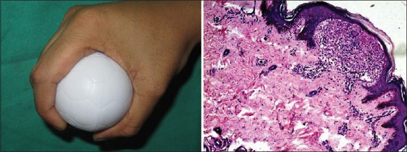
_____ has a characteristic histopathology of "Claw clutching a Ball' appearance
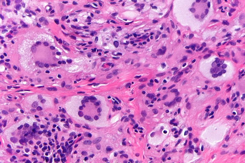
_____ giant cells are lipid-containing macrophages, having a central ring of nuclei while the peripheral cytoplasm is clear due to accumulated lipid
Inflammatory Dermatoses Flashcards for NEET-PG
Study 10 flashcards on Inflammatory Dermatoses for NEET-PG Pathology. These active recall cards cover the key concepts, clinical associations, and high-yield facts from this chapter of Dermatopathology. Each card is designed to test your understanding rather than just recognition, building stronger and more durable memories for exam day.
For personalised spaced repetition scheduling and unlimited flashcards, download the Oncourse app.
Frequently Asked Questions
Are Inflammatory Dermatoses flashcards free?
How many flashcards does this chapter have?
How should I use these flashcards for NEET-PG?
Are there more flashcards for Dermatopathology?
Want unlimited flashcards?
Get full access to all flashcards, spaced repetition, and progress tracking.
Scan to download app