
Anatomy of Visual Pathways — Flashcards

Lesion to areas after the optic chiasm result in _____-ocular and homonymous contra-lateral deficits in vision
K1, K4, and K6 receive _____lateral retinal inputs
Which **magnocellular** layer of the lateral geniculate nucleus (LGN) receives **ipsilateral** retinal input?
K3 and K5 receive _____lateral retinal inputs
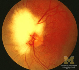
Visual loss is _____ marked in AAION
Pituitary adenoma may present with _____ due to compression of the optic chiasm
The length of the intra-_____ part of the optic nerve is 1mm
_____ is a condition of abnormal myelination of the nerve fibers of the retina, where myelination continues beyond lamina cribrosa and spreads into the retina beyond the optic disc.
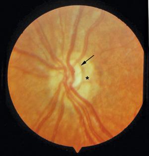
_____ sign is seen in optic nerve hypoplasia
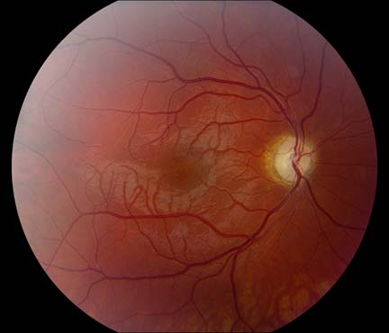
Hyperemic optic disc with telangiectatic (non-leaky on FFA) capillaries is suggestive of _____.
Anatomy of Visual Pathways Flashcards for NEET-PG
Study 10 flashcards on Anatomy of Visual Pathways for NEET-PG Ophthalmology. These active recall cards cover the key concepts, clinical associations, and high-yield facts from this chapter of Neuro-Ophthalmology. Each card is designed to test your understanding rather than just recognition, building stronger and more durable memories for exam day.
For personalised spaced repetition scheduling and unlimited flashcards, download the Oncourse app.
Frequently Asked Questions
Are Anatomy of Visual Pathways flashcards free?
How many flashcards does this chapter have?
How should I use these flashcards for NEET-PG?
Are there more flashcards for Neuro-Ophthalmology?
Want unlimited flashcards?
Get full access to all flashcards, spaced repetition, and progress tracking.
Scan to download app