
Digestive System Histology — Flashcards

Parietal peritoneum is lined by _____ epithelium.
Within which part of the gastric gland are the mucous cells located?_____
The mucosa of the gut wall consists of three layers: an _____, lamina propria, and muscularis mucosa
The _____ cells (chief or parietal) of the stomach stain baso-philic
All three parts of the small intestine contain _____ and microvilli, which increase absorptive surface area
Centroacinar cells are mainly present in the _____.
Plasma cells have abundant RER and a well-developed _____
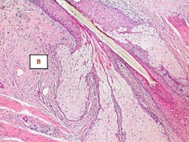
The histopathological image represents the _____ gland, which is a type of holocrine gland.
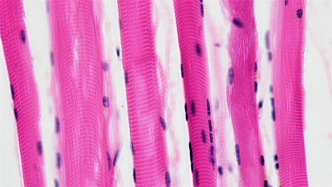
What type of muscle fibre is this? _____
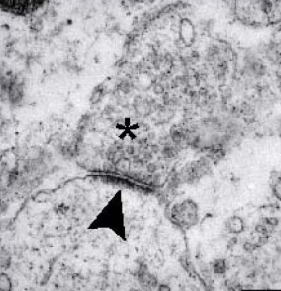
The given image of Electron micrograph shows Presynaptic _____
Digestive System Histology Flashcards for NEET-PG
Study 10 flashcards on Digestive System Histology for NEET-PG Anatomy. These active recall cards cover the key concepts, clinical associations, and high-yield facts from this chapter of Histology. Each card is designed to test your understanding rather than just recognition, building stronger and more durable memories for exam day.
For personalised spaced repetition scheduling and unlimited flashcards, download the Oncourse app.
Frequently Asked Questions
Are Digestive System Histology flashcards free?
How many flashcards does this chapter have?
How should I use these flashcards for NEET-PG?
Are there more flashcards for Histology?
Want unlimited flashcards?
Get full access to all flashcards, spaced repetition, and progress tracking.
Scan to download app