Back
INICET 2026 Pathology Glossary: High-Yield Terms, Tumor Markers and Staining Mnemonics
Master INICET 2026 pathology with this comprehensive glossary of high-yield terms, essential tumor markers (AFP, CEA, PSA, CA125), and staining mnemonics. Includes quick reference tables and study tips.

INICET 2026 Pathology Glossary: High-Yield Terms, Tumor Markers and Staining Mnemonics
Pathology forms the backbone of medical diagnosis and constitutes approximately 15-20% of the INICET (Institute of National Importance Combined Entrance Test) question paper. With over 25,000 candidates competing for limited seats in 2026, mastering pathological concepts, tumor markers, and staining techniques is crucial for success. This comprehensive glossary consolidates the most high-yield pathology terms, essential tumor markers, and memorable staining mnemonics that frequently appear in INICET examinations.
Essential High-Yield Pathology Terms for INICET 2026
Cellular Pathology Fundamentals
Anaplasia: Loss of cellular differentiation and structural organization. Characterized by pleomorphism, abnormal nuclear morphology, and increased mitotic activity. Key feature of malignant tumors. Dysplasia: Abnormal cellular development with loss of uniformity and orientation. Unlike anaplasia, dysplasia is potentially reversible and confined to epithelial tissues. Metaplasia: Replacement of one adult cell type with another adult cell type. Common examples include Barrett's esophagus (squamous to columnar epithelium) and smoker's lung (ciliated columnar to squamous epithelium). Hyperplasia: Increase in cell number leading to organ enlargement. Examples include benign prostatic hyperplasia and endometrial hyperplasia. Hypertrophy: Increase in cell size without increase in cell number. Commonly seen in cardiac muscle under increased workload. Atrophy: Decrease in cell size and number, leading to tissue shrinkage. Can be physiological (thymus in adults) or pathological (muscle wasting in malnutrition).
Inflammation and Healing
Acute Inflammation: Rapid-onset vascular and cellular response characterized by vasodilation, increased vascular permeability, and neutrophil infiltration. Classic signs include rubor (redness), tumor (swelling), calor (heat), dolor (pain), and functio laesa (loss of function). Chronic Inflammation: Prolonged inflammatory response characterized by lymphocytes, macrophages, and plasma cell infiltration. Associated with tissue destruction and repair. Granulomatous Inflammation: Specialized form of chronic inflammation characterized by epithelioid cells, giant cells, and surrounding lymphocytes. Seen in tuberculosis, sarcoidosis, and Crohn's disease. Suppuration: Formation of pus consisting of neutrophils, necrotic tissue, and bacteria. Characteristic of pyogenic bacterial infections.
Neoplasia Terminology
Neoplasm: Abnormal mass of tissue resulting from autonomous proliferation of cells. Can be benign or malignant. Carcinoma: Malignant neoplasm of epithelial origin. Further classified as squamous cell carcinoma, adenocarcinoma, or undifferentiated carcinoma. Sarcoma: Malignant neoplasm of mesenchymal origin including bone, muscle, fat, and connective tissue. Lymphoma: Malignant neoplasm of lymphoid tissue. Broadly classified as Hodgkin's and non-Hodgkin's lymphoma. Carcinoma in Situ: Malignant epithelial cells confined to the epithelial layer without basement membrane invasion. Invasive Carcinoma: Malignant epithelial cells that have breached the basement membrane and invaded underlying tissues.
Comprehensive Tumor Markers Guide
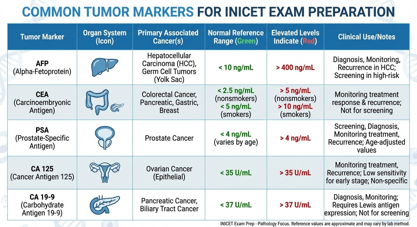
Understanding tumor markers is essential for INICET success, as they frequently appear in multiple-choice questions and clinical scenarios. Here's a comprehensive breakdown of high-yield tumor markers:
Alpha-Fetoprotein (AFP)
Normal Range: <10 ng/mL Elevated in:
Hepatocellular carcinoma (primary liver cancer)
Yolk sac tumors (endodermal sinus tumors)
Embryonal carcinoma
Hepatoblastoma
Neural tube defects (during pregnancy)
Clinical Significance: Most useful for monitoring hepatocellular carcinoma recurrence and treatment response.
Carcinoembryonic Antigen (CEA)
Normal Range: <3 ng/mL (non-smokers), <5 ng/mL (smokers) Elevated in:
Colorectal carcinoma
Pancreatic adenocarcinoma
Gastric carcinoma
Breast carcinoma
Lung adenocarcinoma
Clinical Significance: Primarily used for monitoring colorectal cancer recurrence rather than initial diagnosis.
Prostate-Specific Antigen (PSA)
Normal Range: <4 ng/mL (varies with age) Elevated in:
Prostate adenocarcinoma
Benign prostatic hyperplasia
Prostatitis
Recent prostate manipulation
Clinical Significance: Used for prostate cancer screening and monitoring treatment response.
CA 125 (Cancer Antigen 125)
Normal Range: <35 U/mL Elevated in:
Ovarian carcinoma (especially serous type)
Endometrial carcinoma
Pancreatic carcinoma
Liver disease
Endometriosis
Clinical Significance: Most useful for monitoring ovarian cancer treatment response and detecting recurrence.
CA 19-9 (Cancer Antigen 19-9)
Normal Range: <37 U/mL Elevated in:
Pancreatic adenocarcinoma
Cholangiocarcinoma
Gastric carcinoma
Colorectal carcinoma
Benign biliary obstruction
Clinical Significance: Highest sensitivity for pancreatic adenocarcinoma but not specific.
Human Chorionic Gonadotropin (hCG)
Normal Range: <2 mIU/mL (non-pregnant) Elevated in:
Choriocarcinoma
Hydatidiform mole
Embryonal carcinoma
Yolk sac tumors
Pregnancy
Clinical Significance: Essential for monitoring gestational trophoblastic disease and testicular tumors.
Calcitonin
Normal Range: <10 pg/mL Elevated in:
Medullary thyroid carcinoma
C-cell hyperplasia
Chronic kidney disease
Neuroendocrine tumors
Clinical Significance: Specific marker for medullary thyroid carcinoma and useful for family screening in MEN syndromes.
For comprehensive preparation of tumor markers and their clinical applications, explore our tumor markers lessons and practice with tumor markers MCQs.
High-Yield Staining Techniques and Mnemonics
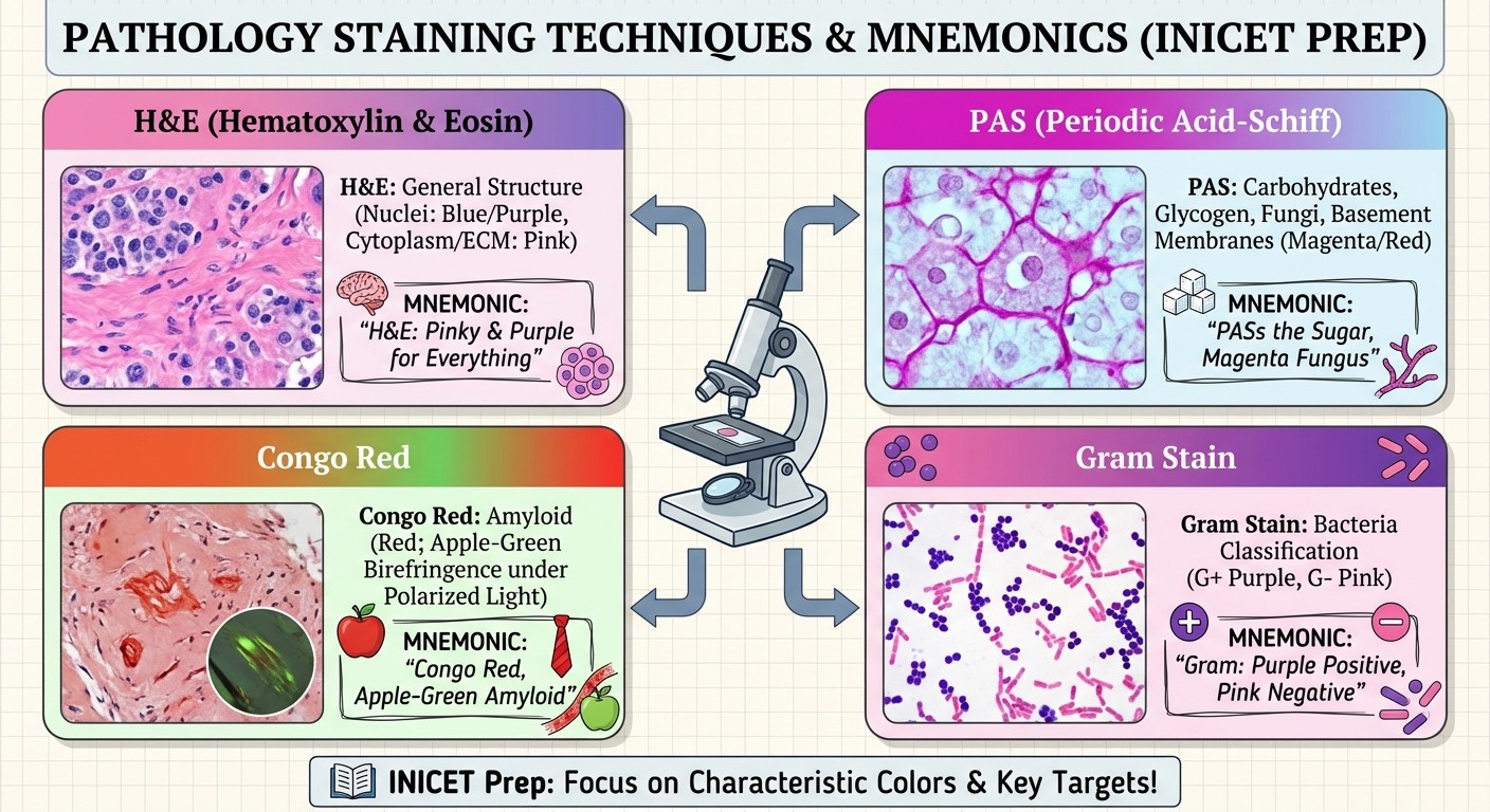
Mastering staining techniques is crucial for INICET pathology questions. Here are the most important stains with memorable mnemonics:
Hematoxylin and Eosin (H&E) Staining
Mnemonic: "Hematoxylin = Heavenly Blue, Eosin = Earthly Pink"
Hematoxylin: Stains nuclei blue/purple
Eosin: Stains cytoplasm pink/red
Uses: Routine histological examination, most common stain
Periodic Acid-Schiff (PAS) Staining
Mnemonic: "PAS = Pink And Sweet"
Stains: Magenta/pink
Targets: Glycogen, mucin, basement membranes, fungal cell walls
Uses: Diagnosing glycogen storage diseases, identifying fungi (Candida, Aspergillus)
Congo Red Staining
Mnemonic: "Congo Red shows Apple Green under Polarized light"
Normal light: Red/pink color
Polarized light: Apple-green birefringence
Targets: Amyloid deposits
Uses: Diagnosing amyloidosis
Gram Staining
Mnemonic: "Gram Positive = Purple, Gram Negative = Pink"
Gram-positive: Retain crystal violet (purple/blue)
Gram-negative: Take up safranin (pink/red)
Uses: Bacterial classification and identification
Acid-Fast Staining (Ziehl-Neelsen)
Mnemonic: "Acid-Fast = Red Fast"
Acid-fast organisms: Red/pink (Mycobacterium, Nocardia)
Non-acid-fast: Blue (background)
Uses: Diagnosing tuberculosis and atypical mycobacterial infections
Reticulin Staining
Mnemonic: "Reticulin = Black Networks"
Stains: Black/dark brown
Targets: Reticular fibers (type III collagen)
Uses: Assessing liver architecture, identifying reticulin collapse
Trichrome Staining (Masson's)
Mnemonic: "Trichrome = Triple colors - Red muscle, Blue collagen, Dark nuclei"
Muscle fibers: Red
Collagen: Blue/green
Nuclei: Dark blue/black
Uses: Distinguishing muscle from connective tissue, assessing fibrosis
Von Kossa Staining
Mnemonic: "Von Kossa = Black Calcium"
Stains: Black/brown
Targets: Calcium deposits and phosphates
Uses: Identifying calcification in tissues
Oil Red O Staining
Mnemonic: "Oil Red = Red Lipids"
Stains: Red/orange
Targets: Neutral fats and lipids
Uses: Diagnosing fatty liver disease, lipid storage disorders
Immunohistochemistry (IHC) Staining
Common Markers and Mnemonics:
Cytokeratin: "CK = Carcinoma Key" (epithelial tumors)
Vimentin: "Vim = Mesenchymal Marker" (sarcomas)
S-100: "S-100 = Schwann cells, Melanocytes, Nerve tissue"
CD20: "CD20 = B lymphocytes"
CD3: "CD3 = T lymphocytes"
For detailed study of staining techniques and their applications, check out our immunologic laboratory techniques lessons and practice with related questions.
Quick Reference Tables for INICET 2026
Tumor Marker Quick Reference
Tumor Marker | Primary Cancer | Normal Range | Key Points |
|---|---|---|---|
AFP | Hepatocellular, Yolk sac | <10 ng/mL | Monitor HCC recurrence |
CEA | Colorectal | <3 ng/mL | Monitor CRC recurrence |
PSA | Prostate | <4 ng/mL | Screening and monitoring |
CA 125 | Ovarian | <35 U/mL | Serous ovarian cancer |
CA 19-9 | Pancreatic | <37 U/mL | Highest for pancreatic adenocarcinoma |
hCG | Gestational trophoblastic | <2 mIU/mL | Monitor GTD and testicular tumors |
Calcitonin | Medullary thyroid | <10 pg/mL | MEN syndrome screening |
Staining Quick Reference
Stain | Color | Target | Primary Use |
|---|---|---|---|
H&E | Blue nuclei, Pink cytoplasm | General morphology | Routine histology |
PAS | Magenta/Pink | Glycogen, mucin, fungi | Glycogen storage, fungal infections |
Congo Red | Red (Apple-green under polarized light) | Amyloid | Amyloidosis diagnosis |
Gram | Purple (positive), Pink (negative) | Bacterial cell wall | Bacterial classification |
Acid-Fast | Red | Mycobacteria | Tuberculosis diagnosis |
Reticulin | Black | Reticular fibers | Liver architecture |
Trichrome | Red muscle, Blue collagen | Connective tissue | Fibrosis assessment |
Advanced Pathology Concepts for INICET
Molecular Pathology Terms
Oncogenes: Genes that promote cell growth and division. When mutated or overexpressed, contribute to cancer development. Examples include MYC, RAS, and HER2. Tumor Suppressor Genes: Genes that normally prevent cell proliferation. Loss of function leads to cancer. Examples include p53 ("guardian of the genome") and Rb (retinoblastoma gene). Proto-oncogenes: Normal genes that can become oncogenes when mutated. They regulate normal cell growth and differentiation. Microsatellite Instability (MSI): Genetic instability caused by defective DNA mismatch repair. Associated with hereditary nonpolyposis colorectal cancer (HNPCC).
Immunopathology Essentials
Type I Hypersensitivity: IgE-mediated immediate hypersensitivity reaction. Examples include anaphylaxis, allergic asthma, and atopic dermatitis. Type II Hypersensitivity: Antibody-mediated cytotoxic reactions. Examples include hemolytic transfusion reactions and autoimmune hemolytic anemia. Type III Hypersensitivity: Immune complex-mediated reactions. Examples include serum sickness and systemic lupus erythematosus. Type IV Hypersensitivity: T-cell mediated delayed-type hypersensitivity. Examples include contact dermatitis and tuberculin skin test.
For comprehensive understanding of immunopathology concepts, explore our immunopathology lessons and reinforce learning with immunopathology flashcards.
Study Tips for INICET 2026 Pathology Success
Memory Techniques
1. Visual Association: Create mental images linking pathological processes with their characteristic features
2. Acronym Formation: Develop memorable acronyms for complex lists (like the tumor markers above)
3. Clinical Correlation: Always connect pathological findings with clinical presentations
4. Case-Based Learning: Practice with clinical vignettes that integrate multiple pathology concepts
High-Yield Topics to Prioritize
Based on previous INICET patterns, focus extra attention on:
Neoplasia (20-25% of pathology questions)
Inflammation and healing (15-20%)
Cardiovascular pathology (10-15%)
Respiratory pathology (10-15%)
Gastrointestinal pathology (10-12%)
Hematopathology (8-10%)
Practice Strategy
1. Daily Review: Spend 30 minutes daily reviewing pathology terms and concepts
2. Weekly Mock Tests: Take pathology-focused mock tests to identify weak areas
3. Image Recognition: Practice identifying pathological specimens and staining patterns
4. Case Discussions: Participate in group discussions of challenging pathology cases
To enhance your pathology preparation, practice with our comprehensive pathology question bank and use our pathology flashcards for effective spaced repetition.
Conclusion: Master Pathology for INICET 2026 Success
Pathology success in INICET 2026 requires systematic understanding of fundamental concepts, memorization of key tumor markers, and mastery of staining techniques. This glossary provides the essential foundation, but consistent practice and application of these concepts in clinical scenarios will determine your success.
Remember that pathology questions in INICET often integrate multiple concepts - a single question might combine tumor markers, staining techniques, and morphological features. Regular practice with high-quality question banks and comprehensive study materials is crucial for developing the pattern recognition skills needed for rapid, accurate answers.
Start your comprehensive pathology preparation today with Oncourse's expertly crafted neoplasia lessons, covering all essential topics from tumor markers to molecular pathology. Our platform offers over 4,000 pathology questions and 10,000+ flashcards designed specifically for Indian medical PG exams, with detailed explanations and memory aids to accelerate your learning.
With systematic preparation using this glossary as your foundation, combined with regular practice and review, you'll be well-equipped to excel in the pathology section of INICET 2026. Success in pathology isn't just about memorization - it's about understanding the underlying principles that govern disease processes and their diagnostic markers.