How to Study Radiology for NEET PG 2026: X-Ray Spotters, CT Signs, High-Yield Topics and Image-Based MCQ Strategy
Master radiology for NEET PG 2026 with this complete guide to X-ray spotters, CT signs, and image-based MCQ strategy. Learn pattern recognition, high-yield topics, and speed training techniques.
How to Study Radiology for NEET PG 2026: X-Ray Spotters, CT Signs, High-Yield Topics and Image-Based MCQ Strategy
You probably stare at a chest X-ray and see nothing but shadows. Meanwhile, that topper from your batch spots a pneumothorax in 3 seconds flat. Radiology carries 15-18 questions in NEET PG — that's 72 marks you cant afford to guess on. The catch? 90% of radiology questions are image-based, testing pattern recognition more than theory.
Here's what separates the scorers from the strugglers: they dont memorize textbook descriptions of findings. They train their eyes to spot abnormalities instantly, like a radiologist would. You have 63 seconds per question — no time to analyze shadows methodically.
This guide breaks down the exact system toppers use to master radiology spotters, high-yield CT signs, and image-based MCQ strategy for NEET PG 2026.
Why Radiology Questions Feel Impossible (And How to Fix That)
NEET PG radiology isnt testing your ability to recite the definition of pneumoperitoneum. It shows you an abdominal X-ray with free air under the diaphragm and asks what you see.
The problem: medical school teaches radiology through textbooks. You memorize that "pneumoperitoneum appears as crescentic radiolucency under the diaphragm" but never actually see 50 different examples of it.
The solution: pattern recognition training. Your brain needs to see the same finding in multiple images until recognition becomes automatic. When you've seen 30 different chest X-rays with pneumothorax, question 31 becomes obvious.
The 4-Phase Radiology Study System
Phase 1: Master Normal Anatomy (Weeks 1-2)
Before spotting abnormalities, you need normal anatomy burned into your visual memory. Start with these high-yield normal films:
Chest X-ray normals:
PA view: cardiac silhouette, lung fields, costophrenic angles, hilar shadows
Lateral view: retrosternal space, vertebral bodies, diaphragm domes
Abdominal X-ray normals:
Gas patterns in bowel
Psoas shadows
Kidney outlines
Pelvic bone landmarks
Spend 15 minutes daily just staring at normal films. Label every structure you can identify. Your eye needs this baseline before it can catch deviations.
Use our comprehensive radiology lessons for systematic coverage of normal anatomy across all imaging modalities.
Phase 2: High-Yield Abnormality Spotters (Weeks 3-6)
Now comes the pattern recognition training. Focus on these guaranteed NEET PG spotters:
Chest X-ray must-knows:
Pneumothorax (collapsed lung edge)
Pleural effusion (costophrenic angle blunting)
Pneumonia consolidation patterns
Cardiomegaly (cardiothoracic ratio >0.5)
Pneumoperitoneum (free air under diaphragm)
CT brain classics:
Acute stroke (loss of gray-white differentiation)
Subdural vs epidural hematoma shapes
Midline shift indicators
Hydrocephalus (ventricular dilatation)
Abdominal imaging favorites:
Bowel obstruction levels and patterns
Free fluid in pelvis
Kidney stones and hydronephrosis
Gallstones and cholecystitis signs
For each finding, see minimum 20 different examples. The key is volume — your pattern recognition improves with repetition, not analysis.
Phase 3: CT and MRI Signs Memorization (Weeks 7-8)
NEET PG loves testing classic radiological signs. These carry easy marks if you know them:
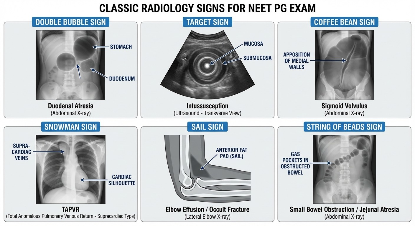
Guaranteed high-yield signs:
Sign | Finding | Condition |
|---|---|---|
Double bubble | Stomach + duodenal bubbles | Duodenal atresia |
Target sign | Concentric rings | Intussusception |
Coffee bean | Dilated sigmoid loop | Sigmoid volvulus |
Snowman | Figure-8 cardiac shadow | TAPVR |
Sail sign | Elevated fat pad | Elbow fracture |
String of beads | Jejunal air-fluid levels | Jejunal atresia |
Hampton's hump | Pleural-based opacity | Pulmonary embolism |
Westermark sign | Regional oligemia | Pulmonary embolism |
Create flashcards for each sign with the image on front, condition on back. The mnemonic I used: "Double Target Coffee brings Snow to Sail String Hampton West" — weird but it stuck.
Practice with our radiology flashcards to reinforce these patterns through spaced repetition.
Phase 4: Image-Based MCQ Speed Training (Weeks 9-10)
This phase separates good scorers from toppers. You need to recognize findings in under 10 seconds, leaving 53 seconds for options analysis.
Speed training protocol:
1. Set 10-second timer per image
2. Call out the finding before time runs out
3. No analysis — just pattern recognition
4. If you miss it, see the same finding 5 more times immediately
Most students spend too long analyzing images. In the real exam, if you dont spot the abnormality quickly, move on. Guessing strategically beats wasting 2 minutes on a difficult spotter.
When practicing image-based questions, I'd recommend using Oncourse's Image Rush game — it trains quick pattern recognition through timed medical image identification, which perfectly mimics the exam pressure.
High-Yield Topics by Weightage
Chest Radiology (4-5 questions)
Must-master spotters:
Pneumothorax patterns (tension vs simple)
Consolidation vs atelectasis vs mass
Cardiac silhouette abnormalities
Mediastinal widening causes
Quick recognition tips:
Pneumothorax: look for absent lung markings at periphery
Effusion: meniscus sign at costophrenic angle
Consolidation: air bronchograms within opacity
Mass: well-defined margins, no air bronchograms
Study systematically with our chest radiology lessons for comprehensive coverage.
Neuroradiology (3-4 questions)
CT brain priorities:
Acute stroke findings (loss of differentiation, hypodense areas)
Hematoma locations and shapes
Mass effect and midline shift
Ventricular system abnormalities
MRI brain basics:
T1 vs T2 signal characteristics
FLAIR sequence findings
Ring-enhancing lesions differential
Master these concepts through our neuroradiology lessons with detailed imaging examples.
Abdominal Radiology (3-4 questions)
X-ray must-knows:
Bowel gas patterns (small vs large bowel)
Obstruction levels and causes
Free air detection (erect vs supine views)
Calcifications (gallstones, kidney stones, pancreatic)
CT abdomen focuses:
Appendicitis findings
Pancreatitis complications
Liver lesion characterization
Urinary tract pathology
Musculoskeletal Radiology (2-3 questions)
Fracture identification:
Common fracture patterns by location
Joint dislocations
Bone tumors vs infections
Arthritis patterns
Pediatric considerations:
Growth plate injuries
Non-accidental trauma patterns
Developmental dysplasia hip
Practice with targeted musculoskeletal radiology questions to reinforce pattern recognition.
Image-Based MCQ Strategy
The 4-Step NEET PG Image Analysis
Step 1: Quality check (2 seconds)
Patient position (AP, PA, lateral, decubitus)
Image quality (rotation, penetration, inspiration)
Patient details for context clues
Step 2: Systematic scan (8 seconds)
Follow the same sequence every time
Chest: heart → lungs → bones → soft tissues
Abdomen: bowels → solid organs → bones → free fluid
Step 3: Abnormality identification (5 seconds)
Compare left vs right for asymmetry
Look for obvious masses, air, fluid collections
Check expected anatomical landmarks
Step 4: Options analysis (remaining time)
Match your finding to the options
Use elimination for close calls
Dont second-guess obvious findings
When I was preparing, I'd use Rezzy AI to discuss complex radiology cases and get instant clarification on findings I wasnt sure about. Having an AI tutor that could analyze images alongside you makes pattern recognition training much more efficient.
Common Trap Avoidance
Trap 1: Normal variants mistaken for pathology
Azygos lobe (normal lung variant)
Prominent thymus in children
Normal fat pads at joints
Trap 2: Technical factors masquerading as disease
Rotation causing apparent cardiomegaly
Poor inspiration mimicking pathology
Motion artifacts
Trap 3: Missing the obvious for the subtle
Focusing on small details while missing gross abnormality
Overanalyzing normal findings
Creating Your Radiology Question Bank
Build a personal collection of image-based questions from these sources:
Primary sources:
Previous NEET PG papers (last 10 years)
AIIMS PG radiology questions
PGI entrance radiology sections
Practice platforms:
Use our comprehensive radiology MCQs for systematic practice across all subtopics
Focus on image-heavy question sets
Time yourself: 60 seconds per question maximum
Quality over quantity rule:
Better to solve 20 questions thoroughly (understanding every finding, every option) than 100 questions superficially.
For each wrong answer:
1. Identify what you missed in the image
2. Find 5 similar images showing the same finding
3. Practice until recognition becomes automatic
Memory Techniques for Radiology Signs
The Location-Finding Method
Instead of memorizing lists, create visual associations:
For pneumothorax: Picture a deflated balloon (collapsed lung) with a visible edge For effusion: Imagine water settling at the bottom of a glass (gravity-dependent) For consolidation: Think of a sponge soaked with fluid (air-filled alveoli become fluid-filled)
The Story Method for Complex Signs
Create mini-stories linking signs to conditions:
"The BABY (duodenal atresia) has TWO BUBBLES (double bubble sign) in their tummy because the connection is blocked, like having two separate balloons instead of one long tube."
These silly stories stick better than textbook definitions.
The Comparison Table Technique
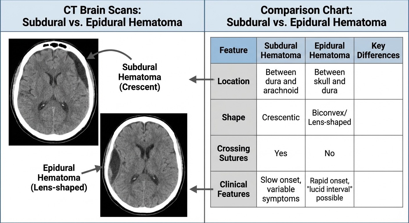
Create comparison tables for commonly confused findings:
Feature | Subdural Hematoma | Epidural Hematoma |
|---|---|---|
Shape | Crescentic | Lens-shaped |
Suture crossing | Crosses sutures | Doesnt cross sutures |
Common location | Cerebral convexity | Temporal region |
Density | Variable | Hyperdense |
Mass effect | Less | More |
Week-by-Week Study Schedule
Weeks 1-2: Foundation Building
Daily time: 45 minutes
Focus: Normal anatomy recognition
Materials: Basic textbook images, normal film libraries
Goal: Identify all normal structures within 30 seconds
Weeks 3-4: Basic Abnormality Recognition
Daily time: 60 minutes
Focus: Common chest and abdominal findings
Materials: Standard abnormality atlases
Goal: Spot obvious abnormalities instantly
Weeks 5-6: Advanced Pattern Recognition
Daily time: 60 minutes
Focus: Subtle findings and differential diagnosis
Materials: NEET PG image banks, case studies
Goal: Differentiate between similar-looking conditions
Weeks 7-8: Speed Training
Daily time: 45 minutes
Focus: Timed practice, quick recognition
Materials: Previous year questions, mock tests
Goal: Complete image analysis within 15 seconds
Weeks 9-10: Exam Simulation
Daily time: 30 minutes
Focus: Full-length practice tests
Materials: Mock exams with image-heavy sections
Goal: Maintain accuracy under time pressure
Technology Tools for Radiology Study
Mobile Apps for Practice
NEET PG radiology apps: For on-the-go practice
Case-based learning apps: Interactive patient scenarios
Anatomy apps: 3D visualization tools
Online Resources
RadiAnt DICOM Viewer: Free software for viewing medical images
Radiopaedia: Comprehensive case database
Teaching file collections: University-shared case libraries
Creating Your Digital Library
Organize saved images by:
1. Body system (chest, abdomen, neuro, MSK) 2. Difficulty level (easy spotters vs subtle findings) 3. Question type (direct identification vs differential diagnosis) 4. Previous mistakes (personal weak areas)
Screenshot every interesting case you encounter. Build your personal radiology atlas over time.
Managing Radiology Anxiety
The Confidence Building Approach
Start with obvious cases where the finding jumps out immediately. Build confidence with easy wins before tackling subtle cases.
Week 1 confidence builders:
Gross pneumothorax with complete lung collapse
Massive pleural effusion with white-out lung
Large pneumoperitoneum with obvious free air
Week 5 challenge cases:
Small apical pneumothorax
Minimal pleural effusion
Trace pneumoperitoneum
The "Good Enough" Principle
You dont need to be a radiologist. You need to score marks. If you can identify the major finding correctly 8 out of 10 times, that's excellent for NEET PG purposes.
Perfect pattern recognition isnt the goal — consistent scoring is.
Common Mistakes and How to Avoid Them
Mistake 1: Trying to Learn Everything
Wrong approach: Attempting to master every possible radiological finding Right approach: Focus on high-yield, commonly tested patterns
The 80/20 rule applies perfectly to radiology. 80% of questions come from 20% of possible findings.
Mistake 2: Pure Memorization Without Context
Wrong approach: Memorizing isolated signs without understanding disease processes Right approach: Connect radiological findings to clinical presentation
Example: Dont just memorize that pneumothorax shows absent lung markings. Understand that a spontaneous pneumothorax typically affects tall, thin young males, and the patient presents with sudden chest pain and dyspnea.
Mistake 3: Neglecting Normal Anatomy
Wrong approach: Jumping straight to pathological findings Right approach: Master normal before tackling abnormal
If you cant identify normal structures confidently, you'll miss obvious abnormalities or worse — call normal findings pathological.
Mistake 4: Analysis Paralysis During Exams
Wrong approach: Spending 3-4 minutes analyzing a single image Right approach: Quick pattern recognition followed by rapid option elimination
Train for speed from day one. Accuracy under time pressure beats perfect analysis that runs out of time.
Frequently Asked Questions
How many radiology questions appear in NEET PG 2026?
Radiology typically carries 15-18 questions in NEET PG, worth approximately 72 marks. About 90% of these are image-based questions testing pattern recognition rather than theoretical knowledge.
Should I buy expensive radiology atlases or use free resources?
Start with free resources like Radiopaedia and previous NEET PG papers. Invest in paid resources only after you've exhausted free materials. The key is volume of practice, not expensive books.
How long does it take to master radiology pattern recognition?
Most students need 8-10 weeks of consistent daily practice to develop reliable pattern recognition skills. However, you can start scoring basic questions within 3-4 weeks of focused study.
What if I cant see anything abnormal in obvious cases?
This usually indicates inadequate normal anatomy knowledge. Go back to studying normal films for 1-2 weeks before attempting pathological cases. You need the baseline before you can spot deviations.
Should I memorize all the radiological signs or focus on common ones?
Focus on the top 20 signs that appear repeatedly in NEET PG. Master these completely rather than knowing 100 signs superficially. The high-yield signs we've listed cover 80% of possible questions.
How important are CT and MRI compared to plain X-rays?
Plain X-rays still dominate NEET PG radiology questions, but CT brain and basic body CT knowledge is essential. MRI questions are less common but appear 2-3 times per exam. Prioritize X-ray → CT → MRI in that order.
Master radiology systematically through consistent practice and pattern recognition training. The visual nature of this subject makes it initially challenging but highly rewarding once you develop the skill.
Prepare smarter with Oncourse AI — adaptive MCQs, spaced repetition, and AI explanations built for NEET PG. Download free on Android and iOS.