How to Ace Image-Based Questions in NEET-PG 2026: Complete Strategy Guide
Master image-based questions in NEET-PG 2026 with proven strategies for radiology, histopathology, anatomy, and clinical photos. Expert tips + practice resources included.
How to Ace Image-Based Questions in NEET-PG 2026: Complete Strategy Guide
Image-based questions constitute 30-40% of the NEET-PG exam, making them one of the highest-scoring yet most challenging components for medical students. Unlike traditional text-based MCQs, these questions demand rapid visual pattern recognition, systematic analytical thinking, and deep subject knowledge across radiology, histopathology, anatomy, and clinical medicine.
In NEET-PG 2026, the National Board of Examinations (NBE) has emphasized competency-based assessment, with image-based questions serving as a critical evaluation tool for clinical reasoning skills. Students who master visual diagnosis often gain a significant competitive advantage, as these questions typically have clear-cut answers once you recognize the pattern.
This comprehensive guide will equip you with proven strategies, systematic approaches, and expert tips to excel in all types of image-based questions in NEET-PG 2026.
Understanding Image-Based Questions in NEET-PG 2026
What Are Image-Based Questions?
Image-based questions in NEET-PG present visual material requiring interpretation, diagnosis, or identification. These questions test your ability to:
Recognize normal and abnormal anatomical structures
Identify pathological changes in tissues and organs
Interpret radiological findings across different imaging modalities
Diagnose clinical conditions from photographs
Correlate gross and microscopic pathology
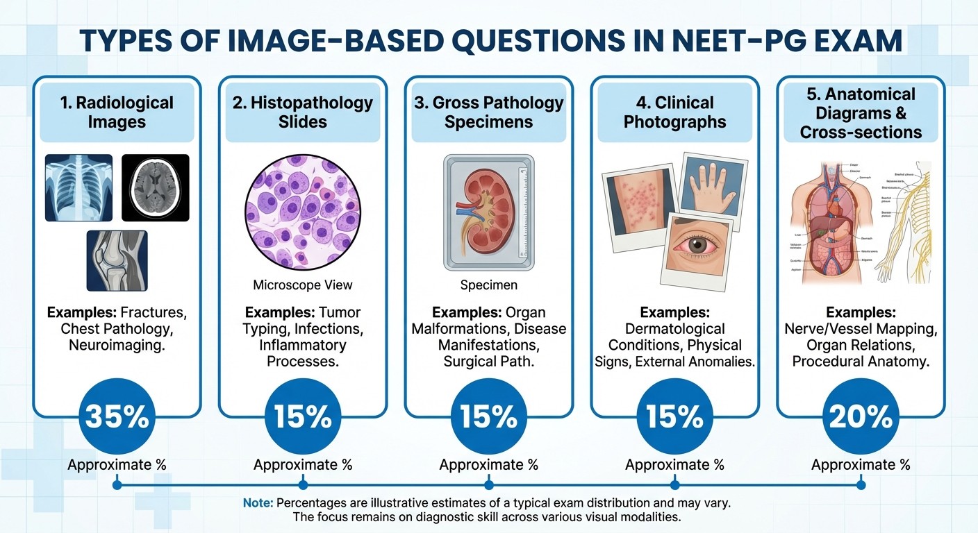
Types of Image-Based Questions in NEET-PG
1. Radiological Images (35-40% of image questions)
X-rays (chest, skeletal, abdominal)
CT scans (brain, chest, abdomen)
MRI images (brain, spine, joints)
Ultrasound images
Nuclear medicine scans
2. Histopathology Slides (25-30%)
H&E stained sections
Special stains identification
Immunohistochemistry patterns
Cytology preparations
3. Gross Pathology Specimens (15-20%)
Organ pathology
Surgical specimens
Autopsy findings
Macroscopic lesions
4. Clinical Photographs (10-15%)
Dermatological conditions
Physical examination findings
Surgical procedures
Clinical signs and symptoms
5. Anatomical Images (10-15%)
Cadaveric specimens
Cross-sectional anatomy
Embryological stages
Anatomical variations
Building Your Foundation: Essential Knowledge Base
Master Normal Anatomy First
Before diving into pathological conditions, ensure rock-solid knowledge of normal anatomy. This forms the foundation for recognizing abnormalities.
High-Yield Normal Anatomy Areas:
Radiographic anatomy of chest: Normal heart size, lung markings, mediastinal contours
Brain CT/MRI anatomy: Ventricular system, major structures, normal variants
Abdominal imaging: Organ positions, normal enhancement patterns
Skeletal landmarks: Age-related changes, normal variants, anatomical positions
Practice with radiological anatomy questions to build your baseline knowledge systematically.
Strengthen Histopathology Recognition
Histopathology questions require immediate pattern recognition of cellular and tissue characteristics.
Key Learning Priorities: 1. Basic tissue types: Epithelial, connective, muscle, nervous tissue patterns 2. Inflammatory patterns: Acute vs. chronic inflammation markers 3. Neoplastic changes: Benign vs. malignant characteristics 4. Organ-specific histology: Liver, kidney, lung, brain, GI tract
Study microscopic anatomy lessons to master tissue identification systematically.
Develop Systematic Pathology Knowledge
Understanding disease processes helps predict what you might see in images.
Essential Pathology Concepts:
Cell injury and death patterns
Inflammation and repair mechanisms
Hemodynamic disorders
Neoplasia classification and grading
Infectious disease manifestations
The RAPID System: Systematic Approach to Image Analysis
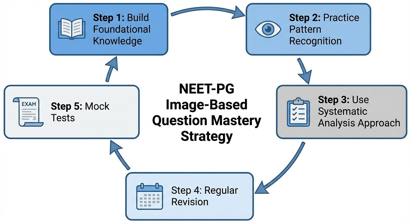
Develop a consistent, systematic approach using the RAPID method:
R - Review the Question Stem
Read the clinical scenario carefully
Note patient demographics (age, sex)
Identify key symptoms or presentation
Look for relevant history or lab values
A - Assess Image Quality and Type
Identify the imaging modality or specimen type
Check image orientation and labeling
Note any artifacts or technical issues
Determine the anatomical region shown
P - Pattern Recognition
Scan for obvious abnormalities first
Compare both sides (if bilateral structures)
Look for symmetry, size, shape, density changes
Note any masses, fluid collections, or structural changes
I - Interpret Findings Systematically
For radiology: Use the systematic approach for each modality
For histopathology: Examine architecture, cell types, special features
For gross pathology: Note size, color, consistency, surface features
For clinical photos: Identify distribution, morphology, associated signs
D - Determine Diagnosis
Correlate imaging findings with clinical presentation
Consider differential diagnoses
Choose the most likely option based on evidence
Eliminate obviously incorrect answers
Subject-Specific Strategies
Mastering Radiology Questions
Chest X-Ray Analysis: 1. Systematic review: Airways → Breathing → Circulation → Diaphragm → Everything else 2. Common patterns to memorize:
- Pneumonia: Air space opacification
- Pneumothorax: Absent lung markings, pleural line
- Pulmonary edema: Bilateral perihilar opacities
- Pleural effusion: Costophrenic angle blunting
CT/MRI Brain: 1. Window settings matter: Bone, brain, blood windows show different pathology 2. Look for symmetry: Compare both hemispheres 3. Check for mass effect: Midline shift, ventricular compression 4. Memorize densities: Blood (hyperdense), CSF (hypodense), gray/white matter
Practice systematically with radiological anatomy questions covering different body systems.
Histopathology Excellence
Low Power First Approach: 1. Architecture assessment: Normal vs. disrupted tissue organization 2. Cellular density: Increased, decreased, or normal cellularity 3. Inflammatory infiltrate: Present or absent, type of cells 4. Special features: Necrosis, hemorrhage, fibrosis High Power Analysis: 1. Cell morphology: Size, shape, nuclear features 2. Nuclear characteristics: Chromatin pattern, nucleoli, pleomorphism 3. Mitotic activity: Number and abnormal forms 4. Cytoplasmic features: Eosinophilic, basophilic, vacuolated
Study microscopic anatomy of different tissues to build pattern recognition skills.
Clinical Photography Analysis
Dermatology Images: 1. Distribution: Localized, generalized, symmetric, asymmetric 2. Morphology: Macule, papule, nodule, vesicle, bulla 3. Color: Erythematous, pigmented, hypopigmented 4. Surface: Smooth, rough, scaling, ulcerated 5. Borders: Well-defined, poorly defined, irregular Physical Examination Findings:
Clubbing: Nail bed angle, fluctuation test
Lymphadenopathy: Size, consistency, mobility
Organomegaly: Hepatosplenomegaly signs
Neurological signs: Specific testing positions
Advanced Study Strategies for 2026
Technology-Enhanced Learning
AI-Powered Practice:
Use AI-driven platforms for adaptive learning
Get instant feedback on image interpretation
Track progress across different image types
Access personalized weak area identification
Oncourse AI platform offers comprehensive image-based question practice with AI-powered explanations and personalized learning paths tailored for NEET-PG 2026.
Spaced Repetition for Visual Memory
Image Flashcard Method:
1. Create digital flashcards with image on front, diagnosis on back
2. Review using spaced repetition algorithms
3. Focus extra time on consistently missed patterns
4. Include both common and rare findings
Practice with anatomy flashcards and pathology flashcards for systematic retention.
Mock Test Strategy
Progressive Difficulty Approach:
Week 1-2: Subject-wise image question practice
Week 3-4: Mixed image questions from multiple subjects
Week 5-6: Full-length tests with time constraints
Week 7-8: Recent pattern questions and image-heavy tests
Common Pitfalls and How to Avoid Them
Time Management Issues:
Problem: Spending too long on single images
Solution: Set 90-second maximum per image question
Practice: Use timer during study sessions
Pattern Recognition Failures:
Problem: Missing obvious findings while looking for rare conditions
Solution: Always check common things first
Approach: "Common things occur commonly" principle
Technical Artifacts Confusion:
Problem: Mistaking technical issues for pathology
Solution: Learn to recognize common artifacts
Examples: Motion artifacts, breathing artifacts, contrast timing
Clinical Correlation Neglect:
Problem: Focusing only on images, ignoring clinical context
Solution: Always correlate findings with patient presentation
Method: Read question stem before examining image
Subject-Wise High-Yield Image Topics
Anatomy (20% of image questions)
Must-Know Areas:
Cross-sectional anatomy: Brain, thorax, abdomen CT levels
Radiological landmarks: Important bony landmarks, soft tissue planes
Embryological images: Key developmental stages
Anatomical variations: Common variants that appear in exams
Study systematically with cross-sectional anatomy lessons.
Pathology (25% of image questions)
Histopathology Priorities:
Inflammation patterns: Acute, chronic, granulomatous
Neoplasia: Benign vs. malignant features, specific tumor types
Organ-specific pathology: Liver, kidney, lung, brain common conditions
Special stains: PAS, reticulin, immunohistochemistry patterns
Gross Pathology Focus:
Cardiovascular: Atherosclerosis, valve disease, cardiomyopathy
Respiratory: Pneumonia, tuberculosis, tumors
GIT: Ulcers, tumors, inflammatory bowel disease
Genitourinary: Kidney stones, tumors, cystic disease
Radiology (30% of image questions)
Chest Imaging:
Common patterns: Pneumonia, tuberculosis, malignancy
Emergency conditions: Pneumothorax, massive PE, aortic dissection
Chronic conditions: COPD, ILD, heart failure
Neuroimaging:
Stroke patterns: Acute, chronic, hemorrhagic vs. ischemic
Tumors: Location-specific features, enhancement patterns
Trauma: Skull fractures, intracranial hemorrhage
Infections: Meningitis, abscess, tuberculoma
Practice with comprehensive radiology question banks covering all major systems.
Clinical Medicine (15% of image questions)
Dermatology:
Infectious: Fungal, bacterial, viral skin infections
Inflammatory: Eczema, psoriasis, autoimmune conditions
Neoplastic: Melanoma, BCC, SCC features
Drug reactions: SJS, TEN, drug eruptions
Physical Signs:
Cardiovascular: Murmur-associated signs, heart failure signs
Respiratory: Clubbing, cyanosis, chest deformities
Neurological: Cranial nerve signs, motor/sensory findings
Endocrine: Thyroid, diabetes, growth hormone effects
Time Management During the Exam
The 90-Second Rule
For image-based questions, allocate maximum 90 seconds per question:
15 seconds: Read question stem and identify image type
45 seconds: Systematic image analysis using RAPID method
20 seconds: Consider differential diagnosis
10 seconds: Select answer and move on
Question Priority System
High Priority (Answer First):
Clear, classic presentations you recognize immediately
Questions with distinctive, pathognomonic findings
Areas of your strongest knowledge base
Medium Priority:
Questions requiring systematic analysis but with clear findings
Differential diagnosis situations with good clinical correlation
Low Priority (Answer Last):
Very subtle findings or rare conditions
Poor quality images with unclear findings
Questions outside your strong subject areas
Final Month Preparation Strategy
Weeks 1-2: Intensive Review
Daily routine: 50 image-based questions across all subjects
Focus: Weak areas identified in previous practice tests
Method: Untimed practice with detailed analysis
Goal: Pattern recognition strengthening
Week 3: Speed Building
Daily routine: 75 image-based questions with time limits
Focus: Applying systematic approach under pressure
Method: Timed practice sessions
Goal: Achieve 90-second average per question
Week 4: Final Polish
Daily routine: Mixed practice tests with image-heavy content
Focus: Maintaining speed while ensuring accuracy
Method: Full-length mock tests
Goal: 80%+ accuracy in image-based questions
Practice your final preparation with comprehensive NEET-PG question banks that include extensive image-based content across all subjects.
Technology Tools and Resources
Essential Apps and Platforms
Image Repository Access:
Radiopaedia: Free radiology image database with detailed explanations
PathologyOutlines: Comprehensive histopathology image collection
DermNet: Extensive dermatology photograph database
Oncourse AI: Integrated image-based question practice with AI feedback
Practice Platforms: 1. Subject-wise practice: Focus on weak areas systematically 2. Mixed question sets: Simulate exam conditions 3. Performance tracking: Monitor improvement over time 4. Doubt resolution: Access expert explanations for missed questions
Creating Your Personal Image Bank
Organize by Categories:
Subject-wise folders (Anatomy, Pathology, Radiology, Medicine)
High-yield vs. rare findings
Frequently missed questions
Last-minute revision images
Review Schedule:
Daily: 20 high-yield images across subjects
Weekly: 50 challenging images from weak areas
Monthly: Complete collection review for retention
Expert Tips from NEET-PG Toppers
Pattern Recognition Mastery
"The more you see, the more you recognize" - Practice 100+ images per topic minimum "Always start with normal" - Master normal anatomy before jumping to pathology "Clinical context is king" - Never ignore the question stem while analyzing images "Time yourself from day one" - Speed comes with practice, not last-minute rushing
Common Mistakes to Avoid
1. Overanalyzing clear-cut cases: Trust your first instinct for obvious patterns
2. Ignoring image quality: Poor images might be testing recognition of limitations
3. Focusing on rare findings: Common conditions appear most frequently
4. Skipping systematic approach: Consistency prevents missing obvious findings
Last-Minute Tips
Day Before Exam:
Review your personal high-yield image collection
Practice 20 quick image questions for confidence
Avoid learning new patterns - stick to revision
Ensure good rest for optimal pattern recognition
During the Exam:
Start with image questions you find easiest to build confidence
Mark uncertain questions for review if time permits
Trust your systematic approach even under pressure
Don't second-guess recognized patterns
Conclusion
Mastering image-based questions in NEET-PG 2026 requires systematic preparation, consistent practice, and strategic time management. The combination of strong foundational knowledge, systematic analytical approach, and extensive practice with varied question types will give you the confidence to excel in this crucial component.
Remember that image-based questions often have the most definitive answers once you recognize the pattern. Your investment in developing visual diagnostic skills will pay dividends not just in NEET-PG but throughout your medical career.
Start implementing these strategies today, maintain consistent practice, and trust in your systematic approach. With dedicated preparation using proven methods, you can transform image-based questions from a challenge into your competitive advantage in NEET-PG 2026.
Ready to start your image-based question mastery journey? Practice with comprehensive image question banks and get AI-powered feedback to accelerate your learning. Your success in NEET-PG 2026 starts with the right preparation strategy implemented consistently.