Functional Imaging Indian Medical PG Practice Questions and MCQs
Practice Indian Medical PG questions for Functional Imaging. These multiple choice questions (MCQs) cover important concepts and help you prepare for your exams.
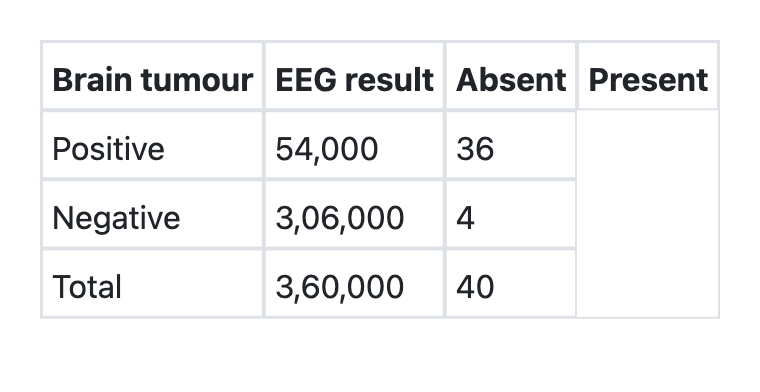
Functional Imaging Indian Medical PG Question 1: What is the sensitivity of EEG for detecting brain tumours as per the information given below?
- A. 90% (Correct Answer)
- B. 99.99%
- C. 0.07%
- D. 85%
Functional Imaging Explanation: ***90%***
- Sensitivity is calculated as **True Positives / (True Positives + False Negatives)**.
- Based on the table provided, among patients with brain tumors (disease positive), 36 cases were correctly identified by EEG and 4 cases were missed.
- Sensitivity = 36/(36+4) = 36/40 = 0.9 or **90%**.
- This indicates that the EEG test correctly identifies 90% of patients who actually have brain tumors.
- High sensitivity is important for screening tests to minimize false negatives.
*99.99%*
- This extremely high percentage is incorrect and not supported by the data.
- It would indicate near-perfect detection of all brain tumor cases, which contradicts the table showing 4 missed cases out of 40.
- Results from miscalculation or misinterpretation of the sensitivity formula.
*0.07%*
- This extremely low value represents a fundamental calculation error.
- Such low sensitivity would indicate the test is essentially useless for detecting brain tumors.
- Does not correspond to any reasonable interpretation of the given data.
*85%*
- While close to the correct answer, this is mathematically incorrect.
- Likely results from calculation error or rounding mistakes.
- The correct calculation (36/40) yields exactly 90%, not 85%.
Functional Imaging Indian Medical PG Question 2: What is the investigation of choice for whole body imaging in metastatic breast cancer?
- A. Angiography
- B. Venography
- C. Magnetic Resonance Imaging
- D. CT Scan (Correct Answer)
Functional Imaging Explanation: ***CT Scan (Correct answer for NEET 2013)***
- **Contrast-enhanced CT scan** was the standard imaging modality for **whole-body staging** in metastatic breast cancer at the time of this exam (2013).
- CT offers **excellent spatial resolution** for detecting metastases in **bone, lung, liver, and lymph nodes**.
- It is widely available, relatively quick, and provides comprehensive anatomical information.
- **Modern Update:** While CT was the standard in 2013, **PET-CT (FDG-PET/CT) is now considered the gold standard** for whole-body staging in metastatic breast cancer due to its combined metabolic and anatomical imaging capabilities. However, PET-CT was not among the options in this historical question.
*Magnetic Resonance Imaging*
- **MRI** is highly sensitive for specific sites, particularly for **brain metastases** and **bone metastases (especially spine and bone marrow)**.
- **Whole-body MRI** protocols are emerging but require longer acquisition times and specialized equipment.
- Not ideal as a single first-line modality for comprehensive whole-body staging compared to CT (or modern PET-CT).
*Angiography*
- **Angiography** is an invasive vascular imaging procedure used to visualize **arterial blood flow**.
- It has **no role in routine metastatic screening or staging** of breast cancer.
- Reserved for specific indications like preoperative vascular mapping or interventional procedures.
*Venography*
- **Venography** specifically visualizes **venous structures** and is used to detect venous thrombosis or venous obstructions.
- It is **not applicable** for detecting solid organ metastases, bone lesions, or lymph node involvement in cancer staging.
Functional Imaging Indian Medical PG Question 3: Which imaging modality is most sensitive for detecting early ischemic stroke?
- A. Ultrasound
- B. PET scan
- C. CT
- D. MRI with DWI (Correct Answer)
Functional Imaging Explanation: ***MRI with DWI***
- **Diffusion-weighted imaging (DWI)** within an MRI scan is highly sensitive in detecting **cytotoxic edema** within minutes of **ischemic stroke** onset. This makes it crucial for early diagnosis and treatment decisions.
- DWI can identify areas of restricted water diffusion, which is a hallmark of acute cellular injury due to **ischemia**, even before changes are visible on conventional T1 or T2-weighted MRI sequences.
*CT*
- While frequently used in acute stroke settings, **non-contrast CT** is primarily used to **rule out hemorrhagic stroke** and may only show subtle or no signs of acute ischemia in the first few hours.
- Early ischemic changes on CT, often referred to as the **"ischemic penumbra"**, may appear hours after stroke onset, making it less sensitive for very early detection compared to DWI.
*Ultrasound*
- **Transcranial Doppler (TCD) ultrasound** can evaluate blood flow velocities in intracranial arteries and detect stenoses or occlusions but is not a primary imaging modality for directly visualizing brain parenchymal ischemia.
- Cervical ultrasound (e.g., **carotid duplex**) assesses extracranial vessels but cannot directly detect **ischemic changes** within the brain tissue itself.
*PET scan*
- **PET (Positron Emission Tomography)** can assess brain metabolism and blood flow but is typically not the preferred or most sensitive modality for **early detection of acute ischemic stroke** due to its complexity, cost, and limited availability in emergency settings.
- PET is more commonly used in research or for assessing chronic conditions and **metabolic abnormalities**, rather than acute stroke diagnosis.
Functional Imaging Indian Medical PG Question 4: Investigation of choice for vascular ring around airway:
- A. PET
- B. Catheter directed angiography
- C. MRI
- D. CT (Correct Answer)
Functional Imaging Explanation: ***CT***
- **CT angiography (CTA)** is the **investigation of choice** for diagnosing vascular rings due to its ability to provide detailed anatomical visualization of the great vessels and their relationship to the trachea and esophagus.
- It offers high spatial resolution, allowing precise identification of the type of vascular anomaly, the degree of **airway and esophageal compression**, and guiding surgical planning.
*PET*
- **PET scans** are primarily used for assessing **metabolic activity**, particularly in oncology or to evaluate organ function, and do not provide sufficient anatomical detail for vascular rings.
- While it can detect metabolically active lesions, it is **not suitable** for visualizing the structural abnormalities of blood vessels and their compressive effects on the airway.
*Catheter directed angiography*
- **Catheter-directed angiography** is an **invasive procedure** involving radiation and contrast, primarily used for assessing blood flow dynamics, identifying stenosis, or guiding interventions.
- While it can visualize vessels, CTA is **less invasive**, provides comparable or superior anatomical detail for vascular rings, and is generally preferred for initial diagnosis.
*MRI*
- **MRI** can provide good soft tissue contrast and visualize vascular structures without radiation, but it is often **less readily available** and can be more challenging for pediatric patients due to the need for sedation and longer scan times.
- For comprehensive anatomical detail including bone and calcifications, and in patients who might struggle with breath-holding, **CT angiography** often offers clearer and more consistent images of complex vascular anatomy.
Functional Imaging Indian Medical PG Question 5: What is the imaging modality of choice for localizing neuroendocrine tumors?
- A. USG
- B. CT
- C. MRI
- D. Somatostatin receptor scintigraphy (Correct Answer)
Functional Imaging Explanation: ***Somatostatin receptor scintigraphy***
- **Somatostatin receptor scintigraphy** is the imaging modality of choice given that most neuroendocrine tumors (NETs) express a high density of somatostatin receptors.
- **68Ga-DOTATATE PET/CT** is the **current preferred technique**, offering superior sensitivity (>90%) and specificity compared to older methods like Indium-111 pentetreotide (Octreoscan).
- This functional imaging allows for **whole-body evaluation** and can detect both primary tumors and metastases, including small lesions that may be missed on conventional anatomical imaging.
- Particularly valuable for detecting occult primary tumors and staging metastatic disease.
*USG*
- **Ultrasound** is useful for initial screening or evaluating superficial NETs, particularly in organs like the pancreas or liver.
- However, its utility is limited by **operator dependence**, gas artifact, and its inability to detect small or deeply located tumors effectively.
- Does not provide functional information about somatostatin receptor expression.
*CT*
- **Computed tomography** provides good anatomical detail and is useful for assessing tumor size, local invasion, and detecting liver metastases.
- While helpful for anatomical characterization, CT can **miss small lesions** (especially <1 cm) and does not provide functional information about receptor status.
- Often used in combination with functional imaging for treatment planning.
*MRI*
- **Magnetic resonance imaging** offers excellent soft tissue contrast and is particularly useful for NETs in the liver and pancreas.
- Superior to CT for detecting liver metastases due to better soft tissue resolution.
- However, MRI has **lower sensitivity for small or widespread lesions** compared to somatostatin receptor imaging and does not provide functional receptor information.
More Functional Imaging Indian Medical PG questions available in the OnCourse app. Practice MCQs, flashcards, and get detailed explanations.