Autoimmune skin diseases: pemphigus vs pemphigoid comparison
Ah, the classic "blistering battle"! Pemphigus vulgaris and Bullous pemphigoid are absolute favorites for examiners because they look somewhat similar but have completely different underlying mechanisms.
Think of it this way: one is a "surface-level" problem (intraepidermal), while the other is a "deep-seated" issue (subepidermal). Let's break down the key differences so you never mix them up again.
The Core Differences
- Pemphigus Vulgaris (PV): This is the "Vicious" one. It involves antibodies against Desmoglein 1 and 3, which are the "glue" holding keratinocytes together. When that glue fails, the cells fall apart (acantholysis), leading to thin, fragile blisters that rupture easily.
- Bullous Pemphigoid (BP): Think of this as "Below" the epidermis. It involves antibodies against BP180 and BP230 in the hemidesmosomes (the "anchors" that tether the epidermis to the basement membrane). Because the entire roof of the blister is the full epidermis, these blisters are tense and much harder to rupture.
I've put together a comparison table to help you visualize these high-yield distinctions side-by-side.
| Feature | Pemphigus Vulgaris (PV) | Bullous Pemphigoid (BP) |
|---|---|---|
| Target Antigen | Desmoglein 1 & 3 (Desmosomes) | BP180 & BP230 (Hemidesmosomes) |
| Blister Level | Intraepidermal (Suprabasal) | Subepidermal |
| Blister Type | Flaccid, fragile, easily ruptured | Tense, firm, less likely to rupture |
| Nikolsky Sign | Positive (Skin sloughs with pressure) | Negative |
| Mucosal Involvement | Common (Often starts in the mouth) | Rare |
| Immunofluorescence | "Fishnet" or "Chicken wire" pattern | Linear pattern along basement membrane |
| Age Group | Younger adults (40-60s) | Elderly (>60s) |
| Prognosis | Potentially fatal if untreated | Generally more benign |
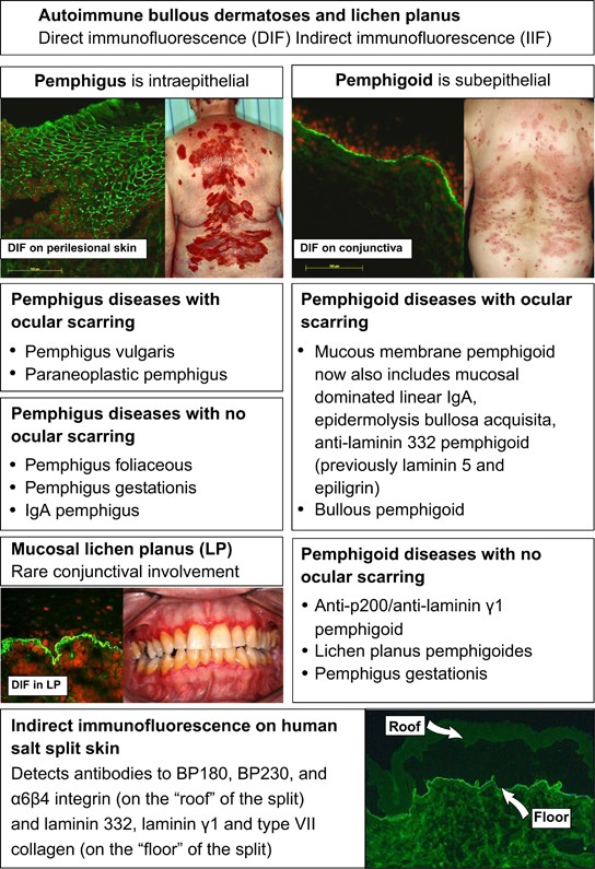
To really nail this for your exams, you need to see the Direct Immunofluorescence (DIF) patterns. They are the "gold standard" for diagnosis and are frequently tested with images.
I'll find some diagrams that show the "fishnet" vs "linear" patterns for you.
This diagram is perfect because it shows you exactly what we were talking about.
Notice the "chicken wire" or "fishnet" pattern in Pemphigus—that's the IgG depositing between the individual keratinocytes. In contrast, the linear pattern in Pemphigoid shows the IgG lining up perfectly along the basement membrane zone.
Mnemonic: Pemphigoid is "B"elow Bullous Pemphigoid = Below the epidermis (subepidermal), BP180/230 antigens, and Blisters are tense!
Would you like to try a few practice questions to see if you can distinguish them in a clinical scenario, or should we look at some flashcards for the high-yield antigens?
Setting up chat...