Ocular anatomy and physiology: aqueous humor, lens and retina
Ocular anatomy is such a fascinating topic—it's basically a high-tech camera system built into your head! Let's dive into the "fluid, the focus, and the film" of the eye: the aqueous humor, the lens, and the retina.
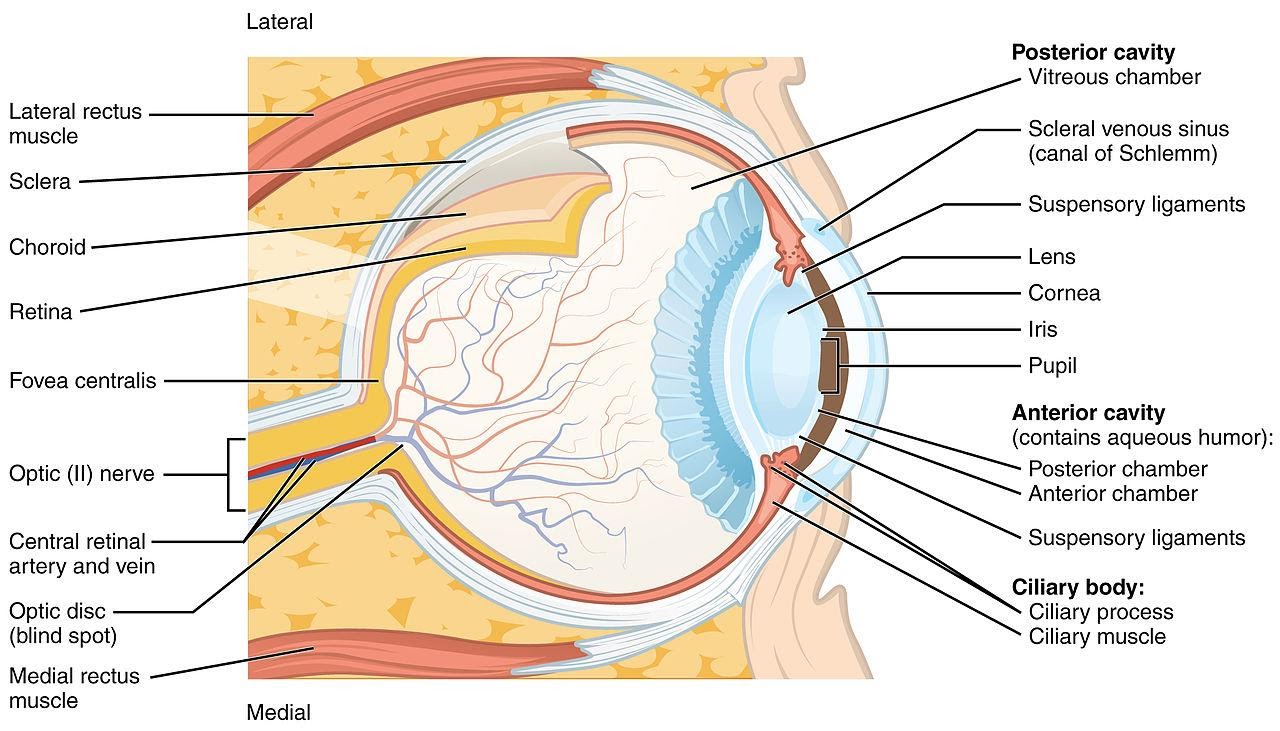
To get us started, I'll pull up a clear diagram of the eye's internal structures so you can visualize where everything sits.
1. Aqueous Humor: The Eye's Internal Plumbing
The aqueous humor is a clear, watery fluid that maintains intraocular pressure (IOP) and provides nutrients to the avascular lens and cornea.
- Production: It's secreted by the ciliary processes in the posterior chamber via active transport (involving carbonic anhydrase).
- Flow: It moves from the posterior chamber, through the pupil, into the anterior chamber.
- Drainage: This is the high-yield part!
- Trabecular Pathway (90%): Through the trabecular meshwork into the Canal of Schlemm.
- Uveoscleral Pathway (10%): Seeps through the ciliary muscle into the suprachoroidal space.
2. The Lens: The Adjustable Focus
The lens is a transparent, biconvex structure that is completely avascular. Its main job is accommodation—changing shape to focus on near objects.
- Structure: It's held in place by zonules (suspensory ligaments) attached to the ciliary body.
- Physiology: When the ciliary muscle contracts, the zonules relax, the lens becomes more spherical (increased power), and you can see that book in front of your face!
I'll create a quick flowchart to help you visualize the aqueous humor pathway, as this is a common exam topic.
That flowchart should help you visualize the flow of aqueous humor, which is key for understanding glaucoma. Now, let's look at the "film" of the camera: the retina.
3. The Retina: The Neural Powerhouse
The retina is where light is converted into electrical signals that the brain can understand. It's composed of 10 distinct layers, which can be a bit of a headache to memorize, but here's the high-yield breakdown:
- Photoreceptors:
- Rods: Responsible for vision in low light (scotopic vision) and peripheral vision. They contain the pigment rhodopsin.
- Cones: Responsible for color vision (photopic vision) and high visual acuity. They are concentrated in the fovea centralis.
- The 10 Layers (Outer to Inner):
- Retinal Pigment Epithelium (RPE)
- Photoreceptor Layer (Rods & Cones)
- External Limiting Membrane (ELM)
- Outer Nuclear Layer (ONL)
- Outer Plexiform Layer (OPL)
- Inner Nuclear Layer (INL)
- Inner Plexiform Layer (IPL)
- Ganglion Cell Layer (GCL)
- Nerve Fiber Layer (NFL)
- Internal Limiting Membrane (ILM)
Mnemonic: "In New Generation, It Is Only Our Education" (Inner to Outer: ILM, NFL, GCL, IPL, INL, OPL, ONL, ELM...) - okay, maybe not the best, but the key is to remember that light travels through the inner layers to reach the photoreceptors at the back!
I'll pull up some lessons and flashcards to help you lock in these anatomical details for your exams.
🗂️ Ocular Physiology Flashcards
Tap to study 25 flashcards in the Oncourse app
📚 Ocular Anatomy Lessons
I've pulled up some great lessons and flashcards that cover everything from the biochemistry of the eye to the detailed anatomy of these structures.
The aqueous humor pathway is particularly high-yield for understanding how different glaucoma medications work (like how beta-blockers decrease production at the ciliary body). The lens is all about that accommodation reflex, and the retina is a masterclass in neural organization.
Would you like to dive deeper into any of these? We could look at the visual cycle (how Vitamin A works in the retina) or maybe the mechanism of accommodation in more detail!
Setting up chat...