Iron deficiency anaemia vs anaemia of chronic disease: blood film and iron studies
This is a classic UKMLA AKT favorite! Distinguishing between Iron Deficiency Anaemia (IDA) and Anaemia of Chronic Disease (ACD) is a bread-and-butter skill for any foundation doctor.
On a blood film, both can actually look quite similar if the ACD has progressed, but there are key nuances:
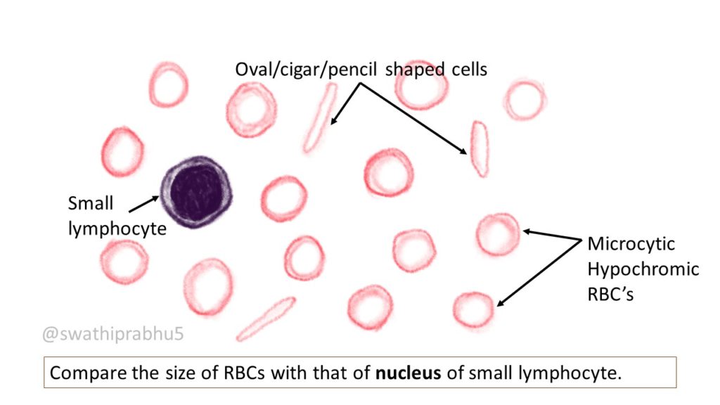
- Iron Deficiency Anaemia (IDA): You'll typically see a microcytic, hypochromic picture. Look for "pencil cells" (elongated RBCs) and occasionally target cells. The cells will have an increased area of central pallor because there's just not enough hemoglobin to fill them up.
- Anaemia of Chronic Disease (ACD): This usually starts as a normocytic, normochromic anaemia. However, in about 30% of long-standing cases (like rheumatoid arthritis or chronic infections), it can become microcytic. You won't usually see the classic "pencil cells" of IDA here.
To make the iron studies easier to memorize, I've put together a comparison table for you.
| Feature | Iron Deficiency Anaemia (IDA) | Anaemia of Chronic Disease (ACD) |
|---|---|---|
| Serum Iron | Low | Low |
| Ferritin | Low (The most sensitive marker) | Normal or High (It's an acute phase reactant) |
| TIBC / Transferrin | High (Body is "hungry" for iron) | Low / Normal (Body is "hiding" iron) |
| Transferrin Saturation | Low | Low / Normal |
| Soluble Transferrin Receptor | High | Normal |
The "golden rule" for the AKT is to look at the Ferritin first. If it's low, it's IDA. If it's normal or high (because it's an acute phase reactant), you're likely looking at ACD.
Let me find a good visual of what those microcytic, hypochromic cells actually look like on a slide.
Take a look at this blood film schematic. It perfectly illustrates the microcytic (small) and hypochromic (pale) nature of the red cells you'd see in IDA.
One last high-yield tip for your AKT prep: if the Ferritin is borderline (because of inflammation), the Soluble Transferrin Receptor (sTfR) can be a real lifesaver. It's elevated in IDA but remains normal in ACD.
It's not always the first-line test, but it's a great "tie-breaker" to have in your back pocket!
Setting up chat...