Bacteriology: gram positive vs gram negative classification and virulence
Bacteriology is the bread and butter of microbiology, and getting the Gram stain classification down is the first step to mastering infectious diseases. It all comes down to that thick (or thin!) peptidoglycan layer in the cell wall.
Let's start with a clear breakdown of the structural differences between these two groups.
The Structural Showdown
The main difference lies in the architecture of their cell walls, which determines how they react to the Gram stain:
- Gram-Positive (+): These have a thick, multi-layered peptidoglycan wall. Think of it like a sturdy brick wall that traps the Crystal Violet stain, making them appear purple under the microscope. They also contain teichoic acids, which help with cell wall stability and mucosal attachment.
- Gram-Negative (-): These have a thin peptidoglycan layer but make up for it with a complex outer membrane. This membrane contains Lipopolysaccharide (LPS). Because the wall is thin, the purple stain washes out during decolorization, and they take up the counterstain (Safranin), appearing pink/red.
I've put together a comparison table to help you visualize these differences side-by-side.
| Feature | Gram-Positive (+) | Gram-Negative (-) |
|---|---|---|
| Color on Gram Stain | Purple / Blue | Pink / Red |
| Peptidoglycan Layer | Thick (multi-layered) | Thin (single-layered) |
| Outer Membrane | Absent | Present |
| LPS (Endotoxin) | Absent | Present |
| Teichoic Acids | Present | Absent |
| Periplasmic Space | Absent | Present |
| Sensitivity to Penicillin | Generally more sensitive | Generally more resistant (due to outer membrane) |
Now, let's talk about Virulence Factors. These are the "weapons" bacteria use to cause disease.
Key Virulence Factors
- Endotoxins (LPS): Found only in Gram-negatives. The Lipid A component of LPS is the toxic part. When the bacteria die and lyse, Lipid A is released, triggering a massive immune response that can lead to septic shock (fever, hypotension, DIC).
- Exotoxins: These are proteins secreted by both Gram-positive and Gram-negative bacteria. Examples include the Tetanus toxin or the Cholera toxin. They are often highly potent and specific in their action.
- Capsules: A sugary coating (polysaccharide) that acts like a "cloak of invisibility," helping bacteria evade phagocytosis by our immune cells.
- Pili/Fimbriae: Hair-like appendages used for attachment to host surfaces.
To help you remember the specific components of the Gram-negative cell wall, which is often a high-yield exam topic, let's look at a diagram.
This diagram perfectly illustrates the "sandwich" structure of the Gram-negative cell wall compared to the "thick wall" of the Gram-positive.
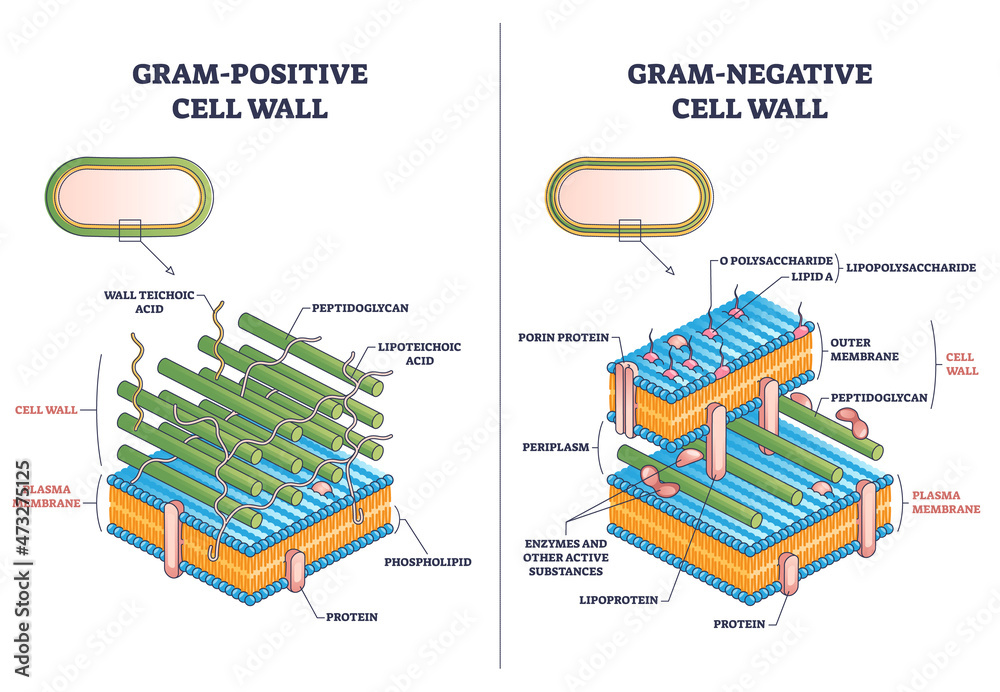
Notice the Lipid A in the Gram-negative outer membrane—that's the "endotoxin" that causes so much trouble in sepsis.
To wrap this up, I've found some great lessons and flashcards that will help you lock in these concepts for your exams.
📚 Bacteriology Lessons
Those lessons cover the taxonomy, structure, and virulence factors in detail. To help you memorize the key differences, I've also pulled up some flashcards.
🗂️ Bacteriology Flashcards
Tap to study 10 flashcards in the Oncourse app
Those flashcards are perfect for a quick review of the cell wall differences and the distinction between exotoxins and endotoxins.
Remember, the key to mastering bacteriology is to always link the structure (like the Gram-negative outer membrane) to the clinical presentation (like septic shock from Lipid A).
Is there a specific bacterium or a particular virulence mechanism you'd like to dive deeper into?
Setting up chat...