Diseases of the ear: otitis media, cholesteatoma, hearing loss types
Ah, ENT! The ear can be a bit of a maze (pun intended), but it's super high-yield for exams like NEET-PG and INI-CET. We're looking at everything from common infections to the tricky structural issues like cholesteatoma.
Let's start by getting some solid lessons lined up so you have the full theory at your fingertips.
📚 Ear Disease Lessons
Those lessons cover the heavy lifting, but let's break down the "Big Three" you mentioned so you have a clear mental map.
1. Otitis Media (OM)
This is essentially inflammation of the middle ear. You'll mostly deal with two main types in exams:
- Acute Otitis Media (AOM): Usually follows a URI. Look for a bulging, erythematous tympanic membrane (TM) and severe pain.
- Otitis Media with Effusion (OME): Fluid behind the TM without signs of acute infection. Often called "glue ear" in kids. The TM might look retracted or show air-fluid levels.
2. Cholesteatoma
Think of this as "skin in the wrong place." It's a non-neoplastic keratinizing squamous epithelium collection in the middle ear or mastoid.
- Key Sign: A pearly white mass behind the TM or in a retraction pocket (usually in the pars flaccida).
- Danger: It’s bone-eroding! It can eat through the ossicles (causing hearing loss) or even the tegmen tympani (leading to CNS complications).
3. Hearing Loss Types
This is a classic "bread and butter" topic. I've put together a comparison table to help you distinguish them quickly during a clinical exam or a MCQ.
| Feature | Conductive Hearing Loss (CHL) | Sensorineural Hearing Loss (SNHL) |
|---|---|---|
| Pathology | Problem in External or Middle Ear | Problem in Inner Ear (Cochlea) or CN VIII |
| Common Causes | Wax, Otitis Media, Otosclerosis | Presbycusis, Noise trauma, Ototoxicity |
| Rinne Test | Negative (BC > AC) | Positive (AC > BC) |
| Weber Test | Lateralizes to the Affected ear | Lateralizes to the Healthy ear |
| Audiometry | Air-Bone Gap present | No Air-Bone Gap (both lines drop) |
That table should help you nail those tuning fork questions!
One last thing: Cholesteatoma can be tricky to visualize just from a description. Let me find a good clinical image of it so you can recognize it instantly if it pops up in an image-based question.
That image shows a classic case of middle ear pathology. Notice the pearly white appearance near the pars flaccida—that's your big clue for cholesteatoma.
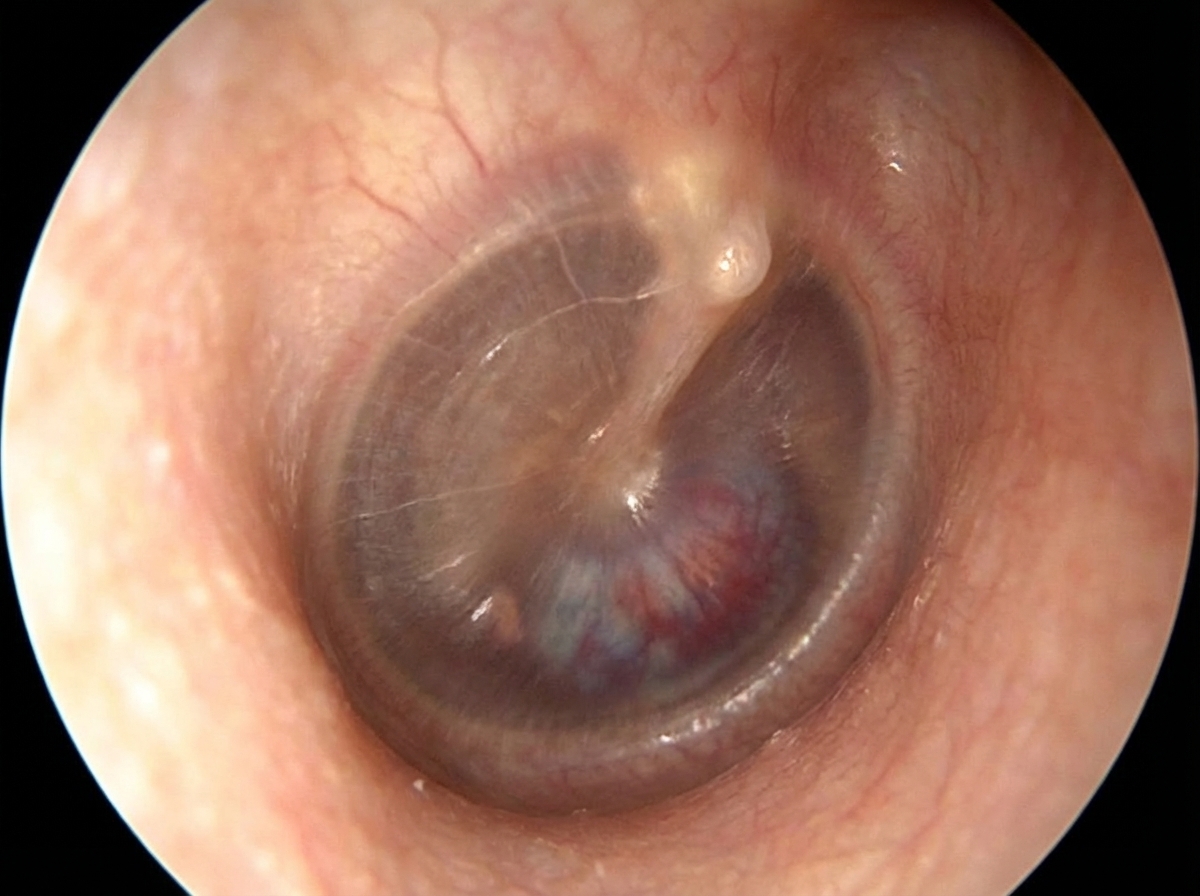
This otoscopic view shows a whitish, keratinous mass in the superior part of the tympanic membrane, which is characteristic of a cholesteatoma. You can also see signs of inflammation and fluid, which often accompany chronic ear disease.
Since these topics are so high-yield, would you like to try a quick quiz to see how well you can differentiate between these conditions in a clinical scenario?
Setting up chat...