Dermatopathology: skin biopsy patterns and histological diagnosis
Dermatopathology can feel like looking at a Rorschach test sometimes, but it’s actually very logical once you break it down into specific histological patterns. Think of it as "pattern recognition" rather than just memorizing every single disease.
I'll start by finding a good visual overview of these biopsy patterns to help you visualize the architecture we're talking about.
While dermoscopy is great for the surface, the real magic happens when we look at the tissue architecture under the microscope. Let me see if I can find a specific histological diagram for you.
That's a great find! This image perfectly captures the "Big Four" patterns you'll encounter in dermatopathology: psoriasiform, spongiotic, vesiculobullous, and lichenoid.
I'll display this for you now so you can see the architectural differences side-by-side.
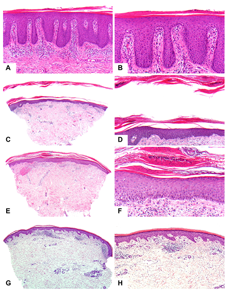
This H&E stained image illustrates the major patterns: Psoriasiform (A, B), Spongiotic (C, D), Vesiculobullous (E, F), and Lichenoid/Interface (G, H).
To make this even easier to digest, I've put together a summary table of these patterns, their key histological features, and the classic diagnoses associated with each.
| Pattern | Key Histological Features | Classic Diagnoses |
|---|---|---|
| Psoriasiform | Regular epidermal hyperplasia (acanthosis), elongated rete ridges, parakeratosis | Psoriasis, Pityriasis rubra pilaris |
| Spongiotic | Intercellular edema (spongiosis), intraepidermal vesicles | Eczema/Dermatitis (allergic, irritant, atopic) |
| Lichenoid (Interface) | Band-like lymphocytic infiltrate at the dermal-epidermal junction, basal cell vacuolar degeneration | Lichen planus, Lupus erythematosus |
| Vesiculobullous | Blister formation (subepidermal or intraepidermal) | Pemphigus vulgaris, Bullous pemphigoid |
| Granulomatous | Collections of epithelioid histiocytes (granulomas) | Sarcoidosis, Tuberculosis, Leprosy |
| Panniculitis | Inflammation of subcutaneous fat (septal or lobular) | Erythema nodosum, Erythema induratum |
This is a great foundation! To really lock this in for your exams, I'll pull up some relevant lessons and flashcards that go deeper into these specific patterns.
📚 Dermatopathology Lessons
Those lessons cover the theory, but flashcards are the best way to test your pattern recognition. Let me find some for you.
🗂️ Dermatopathology Flashcards
Tap to study 24 flashcards in the Oncourse app
Dermatopathology is all about identifying the "neighborhood" (the pattern) before you look for the specific "house" (the diagnosis).
Remember these high-yield histological "buzzwords" for your exams:
- Psoriasis:
Acanthosis,Parakeratosis,Munro's microabscesses. - Lichen Planus:
Saw-tooth rete ridges,Wickham striae(clinically),Civatte bodies. - Eczema:
Spongiosis(intercellular edema). - Pemphigus Vulgaris:
Acantholysis,Tombstone appearance.
You've got the visual guide, the summary table, and plenty of practice material now. Let me know if you want to dive deeper into any of these specific patterns!
Setting up chat...