Dermatopathology: skin biopsy patterns and high-yield histological features
Dermatopathology can feel like looking at a pink and purple abstract painting at first, but once you recognize the specific "patterns" of inflammation and the "signature" cells, it becomes one of the most logical parts of the USMLE!
To get us started, I'll find a clear diagram that breaks down these fundamental biopsy patterns so you can visualize the architecture we're talking about.
That diagram gives a great clinical overview, but let's dive into what you'll actually see under the microscope. Dermatopathology is usually categorized into a few "big" patterns.
I'm going to pull up a more specific histological comparison to help you distinguish between things like Psoriasis and Lichen Planus at the cellular level.
This comparison is high-yield gold for the USMLE! It perfectly illustrates the two most common "interface" and "psoriasiform" patterns you'll be tested on.
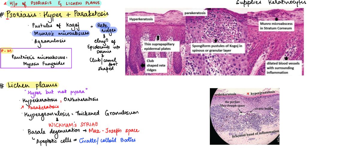
That image really helps visualize the "saw-tooth" appearance of Lichen Planus versus the "test-tube" rete ridges of Psoriasis.
Here's a breakdown of the major patterns you'll need to recognize:
1. Psoriasiform Pattern
Characterized by regular epidermal thickening (acanthosis).
- Classic Example: Psoriasis.
- High-Yield Features:
- Munro microabscesses: Neutrophils in the stratum corneum.
- Spongiform pustules of Kogoj: Neutrophils in the spinous layer.
- Auspitz sign: Pinpoint bleeding when scales are removed (due to thin suprapapillary plates and dilated vessels).
2. Lichenoid (Interface) Pattern
Inflammation that "hugs" the dermo-epidermal junction.
- Classic Example: Lichen Planus.
- High-Yield Features:
- Saw-tooth rete ridges: Jagged appearance of the epidermis.
- Civatte bodies: Apoptotic keratinocytes (pink globules).
- Wickham striae: White lacy lines seen clinically, representing hypergranulosis.
3. Spongiotic Pattern
Intercellular edema (spongiosis) within the epidermis.
- Classic Example: Eczema (Atopic Dermatitis), Contact Dermatitis.
- High-Yield Features: "Fluid" between keratinocytes, making them look like they're held together by tiny bridges (desmosomes).
4. Vesiculobullous Pattern
Blister formation at different levels.
- Pemphigus Vulgaris: Intraepidermal blister (acantholysis) with a "tombstone" appearance of the basal layer.
- Bullous Pemphigoid: Subepidermal blister (tense bullae) with eosinophils.
I've put together a quick reference table for the "buzzword" histological features that often show up in question stems.
| Feature | Associated Condition | Description |
|---|---|---|
| Munro microabscesses | Psoriasis | Neutrophils in the parakeratotic stratum corneum |
| Civatte (Colloid) bodies | Lichen Planus | Apoptotic keratinocytes in the basal layer |
| Pautrier microabscesses | Mycosis Fungoides | Clusters of atypical T-cells in the epidermis |
| Birbeck granules | Langerhans Cell Histiocytosis | "Tennis racket" shaped organelles on EM |
| Cowdry A bodies | HSV / VZV | Eosinophilic intranuclear inclusions |
| Donovan bodies | Granuloma Inguinale | Intracellular safety-pin shaped organisms (Klebsiella) |
| Medlar bodies | Chromoblastomycosis | "Copper penny" fungal structures |
That table covers the most common "buzzwords" you'll see in question stems. To help you solidify these concepts, I'll pull up some relevant lessons and flashcards that focus on these high-yield dermatopathology patterns.
📚 Dermatopathology Lessons
Those lessons on inflammatory dermatoses and blistering diseases are exactly what you need to master these patterns.
To help you drill the "buzzwords" and histological features into your memory, I'll pull up some high-yield flashcards.
🗂️ Dermatopathology Flashcards
Tap to study 29 flashcards in the Oncourse app
Those flashcards are perfect for quick-fire review!
Dermatopathology is all about pattern recognition. Once you can differentiate the "hugging" inflammation of Lichen Planus from the "test-tube" ridges of Psoriasis, you've already mastered a huge chunk of the USMLE dermatology section.
If you're feeling confident, we could try a quick Clinical Rounds game to see how these biopsy findings apply to real patient cases, or I can pull up some Practice Questions to test your knowledge. What do you think?
Setting up chat...