Cardiovascular radiology: CTA, echocardiography and coronary angiography interpretation
Hey there! Diving into cardiovascular radiology is like becoming a detective for the heart. We've got some heavy hitters here: CTA, Echo, and Angiography. Each one gives us a different "view" of what's going wrong, whether it's a plumbing issue (vessels) or a pump issue (valves and muscle).
Let's start with Coronary Computed Tomography Angiography (CCTA). This is our go-to for a non-invasive look at the coronary arteries. It's brilliant for ruling out Coronary Artery Disease (CAD) in patients with low-to-intermediate risk. We're looking for things like the Calcium Score (a predictor of future events) and the degree of luminal stenosis. I'll pull up a clear image of a CTA scan so you can see how the vessels are visualized.
That image shows exactly how we can spot calcified plaques in the coronary arteries using CT. It's a game-changer for risk assessment!
Next up is Echocardiography, the workhorse of cardiology. Whether it's a Transthoracic Echo (TTE) or the more invasive Transesophageal Echo (TEE), we're looking at the heart in motion. Key things to interpret here are the Ejection Fraction (EF) (how well the pump is working), valvular function (stenosis or regurgitation), and wall motion abnormalities (which might hint at an old MI). Let me find a classic "Apical Four-Chamber View" for you.
The apical four-chamber view is like the "home base" for Echo—it gives you a great look at all the chambers and the valves in one shot.
Finally, we have the "Gold Standard": Invasive Coronary Angiography. This is where we actually thread a catheter into the coronaries and inject contrast under fluoroscopy. When interpreting these, we look for the percentage of stenosis and the TIMI flow grade (how fast the blood is moving through the vessel). It's both diagnostic and therapeutic, as we can go straight to angioplasty or stenting if we find a blockage. Let's see if I can find a shot of a vessel with a clear narrowing.
That angiogram shows exactly why it's the gold standard—you can see the narrowing so clearly!
I've put together a visual summary of these three modalities for you below. Take a look at the differences in how they visualize the heart.
Cardiovascular Imaging Modalities
| Modality | Key Visual | Description |
|---|---|---|
| Coronary CTA | 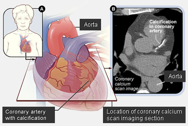 | Shows anatomical detail of the coronary arteries and calcified plaques. |
| Echocardiography |  | Real-time ultrasound showing chambers, valves, and muscle function. |
| Coronary Angiography | 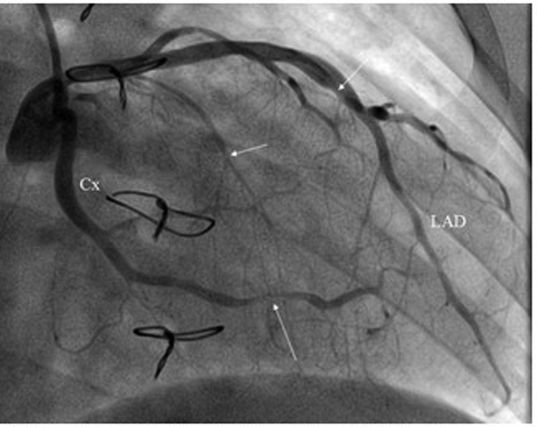 | Invasive fluoroscopy showing luminal narrowing (stenosis) in real-time. |
Seeing them side-by-side really helps clarify when to use which one! CTA for non-invasive screening, Echo for functional assessment, and Angiography for definitive diagnosis and treatment.
To help you master the interpretation of these scans for your exams, I've pulled up some high-yield lessons that break down the specific findings you'll need to know.
📚 Cardiovascular Radiology Lessons
Those lessons will give you the deep dive you need, especially on the specific indications and interpretation criteria for each modality.
If you want to test your knowledge on these, we could jump into a quick @Quiz on cardiac imaging, or I can find some @Flashcards to help you memorize the TIMI flow grades or Echo views. Which one sounds better to you?
Setting up chat...