Brainstem nuclei: locations, functions and clinical correlations
The brainstem is essentially the "Grand Central Station" of the nervous system. It’s packed with nuclei that control everything from your ability to move your eyes to your heart rate. To make this digestible, it's best to break it down by level: Midbrain, Pons, and Medulla.
I'll start by pulling up some high-yield lessons that break these nuclei down by their anatomical levels and functional columns.
📚 Brainstem Nuclei Overview
That's a solid start. To help you visualize where these nuclei sit, I've put together a summary table. A great rule of thumb for the USMLE is the "Rule of 4": 4 nerves in the midbrain (actually 2), 4 in the pons, and 4 in the medulla.
Let's look at the functional breakdown:
| Level | Cranial Nerve Nuclei | Key Functions | Clinical Correlation |
|---|---|---|---|
| Midbrain | CN III (Oculomotor), CN IV (Trochlear) | Eye movement, pupillary constriction (Edinger-Westphal) | Weber Syndrome: CN III palsy + contralateral hemiparesis |
| Pons | CN V (Trigeminal), VI (Abducens), VII (Facial), VIII (Vestibulocochlear) | Facial sensation/mastication, eye abduction, facial expression, hearing/balance | Locked-in Syndrome: Basilar artery stroke affecting ventral pons |
| Medulla | CN IX (Glossopharyngeal), X (Vagus), XI (Accessory), XII (Hypoglossal) | Swallowing, gag reflex, parasympathetic output to viscera, tongue movement | Wallenberg Syndrome (PICA): Lateral medullary syndrome |
Now, to really lock this in for your exams, you need to know the functional columns. For example, the motor nuclei are always medial, and the sensory nuclei are always lateral (separated by the sulcus limitans).
I'll find a diagram that shows this medial-to-lateral organization, as it's a classic USMLE question.
Check out this cross-section of the medulla. It perfectly illustrates how the Hypoglossal nucleus (CN XII) is right in the middle (medial), while the sensory nuclei like the Vestibular nuclei (CN VIII) are pushed out to the side (lateral).
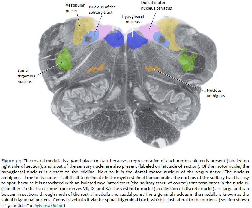
This diagram shows the classic medial-to-lateral organization: Motor nuclei (XII) are medial, while sensory nuclei (Solitary tract, Vestibular) are lateral.
High-Yield Clinical Correlations
-
Medial Medullary Syndrome (ASA Stroke):
- Affects the Hypoglossal nucleus (CN XII).
- Presentation: Tongue deviates toward the side of the lesion, plus contralateral hemiparesis (pyramids) and loss of vibration/proprioception (medial lemniscus).
-
Lateral Medullary Syndrome (Wallenberg - PICA Stroke):
- Affects the Nucleus Ambiguus (CN IX, X, XI) and Vestibular nuclei.
- Presentation: Dysphagia, hoarseness (nucleus ambiguus), and vertigo/nystagmus (vestibular nuclei).
-
MLF (Medial Longitudinal Fasciculus):
- This is the "highway" connecting CN VI in the pons to CN III in the midbrain.
- Lesion: Internuclear Ophthalmoplegia (INO), commonly seen in Multiple Sclerosis.
Would you like to try a few practice questions on these syndromes, or maybe a quick game to test your recall of which nerve belongs to which level?
Setting up chat...