Basic dermatology: primary and secondary skin lesions terminology
Dermatology is often called the "visual specialty" because so much of the diagnosis depends on accurately describing what you see. We divide skin lesions into primary (the initial physical change) and secondary (changes that occur due to evolution, trauma, or infection of a primary lesion).
Let's start with the primary lesions, which are the building blocks of any skin exam.
| Lesion Type | Description | Examples |
|---|---|---|
| Macule | Flat, non-palpable change in color, < 1 cm | Freckles, Petechiae |
| Patch | Flat, non-palpable change in color, > 1 cm | Vitiligo, Melasma |
| Papule | Solid, raised lesion, < 1 cm | Acne, Warts |
| Plaque | Raised, flat-topped lesion, > 1 cm | Psoriasis |
| Nodule | Solid, deep-seated raised lesion, > 1 cm | Lipoma, Erythema nodosum |
| Vesicle | Fluid-filled (serous), < 1 cm | Herpes simplex, Chickenpox |
| Bulla | Fluid-filled (serous), > 1 cm | Bullous pemphigoid |
| Pustule | Pus-filled vesicle | Acne pustule, Folliculitis |
| Wheal | Transient, edematous papule or plaque | Urticaria (Hives) |
Secondary lesions are what happens when a primary lesion is scratched, infected, or simply ages. They are crucial for understanding the chronicity of a skin condition.
| Lesion Type | Description | Examples |
|---|---|---|
| Scale | Flaking or shedding of the stratum corneum | Psoriasis, Dandruff |
| Crust | Dried serum, blood, or pus (a "scab") | Impetigo (honey-colored) |
| Erosion | Loss of part of the epidermis; heals without scarring | Ruptured vesicle |
| Ulcer | Loss of epidermis and part of dermis; heals with scarring | Pressure sore, Syphilis |
| Fissure | Linear crack in the skin | Athlete's foot, Cheilitis |
| Atrophy | Thinning of the skin (parchment-like) | Topical steroid overuse |
| Lichenification | Thickening of skin with accentuated markings from rubbing | Chronic eczema |
| Excoriation | Linear erosion caused by scratching | Scabies, Neurodermatitis |
| Scar | Replacement of normal skin with fibrous tissue | Post-surgical, Keloid |
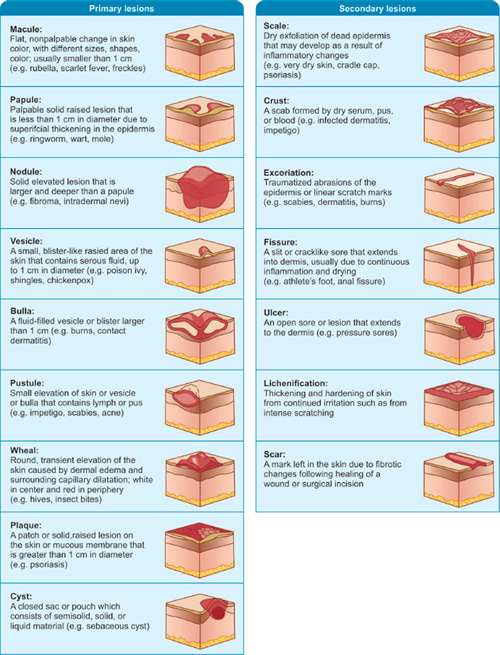
Visualizing these differences is key to mastering dermatology. Let me find a diagram that illustrates these primary and secondary lesions for you.
This diagram provides a clear cross-sectional view of how these lesions sit within the layers of the skin, which is very helpful for understanding their depth.
To dive deeper into how these lesions present in specific diseases, I've pulled up some relevant lessons for you.
📚 Dermatology Morphology Lessons
Mastering these terms is the first step in any dermatology rotation. If you'd like to practice identifying these in clinical scenarios or want to see more specific examples of conditions like psoriasis or impetigo, just let me know!
Setting up chat...