Bacterial skin infections: impetigo, cellulitis, SSSS and management
Bacterial skin infections are a bread-and-butter topic for exams like NEET-PG and INI-CET. They often present as "spotters" or clinical vignettes where the description of the lesion gives away the diagnosis.
Let's dive into the "big three" you asked about!
1. Impetigo
This is the most superficial of the bunch, usually affecting the epidermis. It's highly contagious and common in kids.
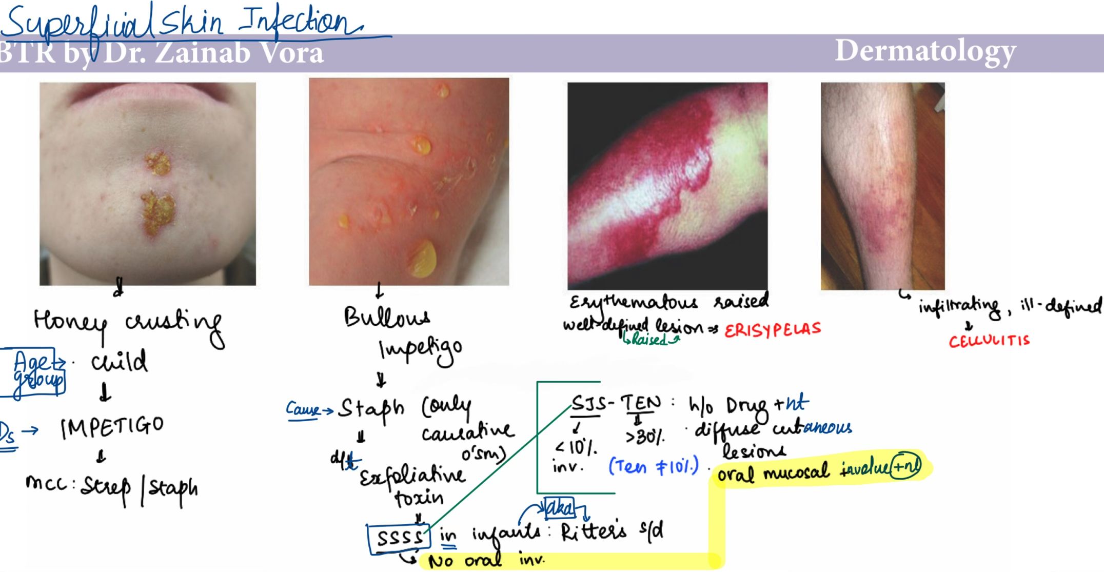
- Non-bullous (70%): Caused by S. aureus or S. pyogenes. Look for the classic =="honey-colored crusts"== on an erythematous base, often around the nose or mouth.
- Bullous: Almost always S. aureus. It produces an exfoliative toxin that causes large, flaccid bullae.
- Management:
- Localized: Topical Mupirocin or Retapamulin.
- Widespread: Oral antibiotics like Cephalexin or Dicloxacillin.
2. Cellulitis vs. Erysipelas
These are deeper infections. The key is knowing how to tell them apart!
- Erysipelas: Superficial dermis and upper lymphatics. It has ==sharply demarcated borders== and is usually caused by S. pyogenes.
- Cellulitis: Deeper dermis and subcutaneous fat. The borders are ill-defined and "spreading." Common causes are S. pyogenes and S. aureus.
- Management:
- Empiric therapy usually targets Gram-positives (Flucloxacillin, Cephalexin).
- If MRSA is suspected, think Vancomycin or Linezolid.
3. Staphylococcal Scalded Skin Syndrome (SSSS)
This is a systemic reaction to the exfoliative toxins (ETA, ETB) produced by certain strains of S. aureus.
- Clinical Features: It starts with a perioral crusting and fever, progressing to widespread erythema and skin peeling.
- Key Sign: ==Nikolsky sign is positive== (the top layer of skin slips off with slight pressure).
- Management:
- Hospitalization is usually required.
- IV antibiotics (Nafcillin, Oxacillin, or Vancomycin).
- Fluid and electrolyte management (similar to burn care).
I'll find some high-yield images to help you visualize these differences.
This image perfectly captures the differences we just discussed. Notice the honey-colored crusts in the first panel and the sharp borders of erysipelas compared to the diffuse redness of cellulitis.
To get into the nitty-gritty details, I've pulled up some relevant lessons for you. After that, we can test your knowledge with a quick quiz!
📚 Bacterial Skin Infections Lessons
Ready to see how much you've retained? I've put together a quiz with some high-yield questions on these bacterial infections.
✍️ Bacterial Skin Infections Quiz
Tap to practice questions in the Oncourse app
You're all set! Those lessons and the quiz should help you master these skin infections. If you have any more questions or want to dive into another topic, just let me know!
Setting up chat...