Autonomic nervous system anatomy: sympathetic vs parasympathetic pathways
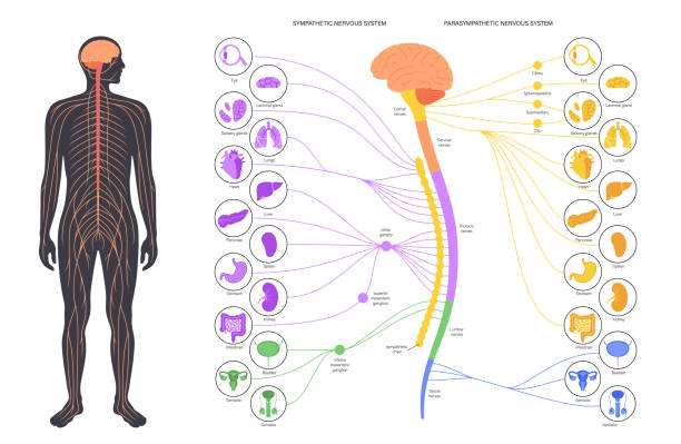
The Autonomic Nervous System (ANS) is basically the body's "autopilot," and keeping the sympathetic and parasympathetic pathways straight is a classic med school hurdle. Let's start with a visual to see how they're laid out differently across the spinal cord and organs.
That diagram gives a great overview of how the two systems are physically organized. To make the high-yield anatomical differences easier to digest, I've put together a quick comparison table for you.
The key to remembering these is the "origin" and the "length" of the fibers. Here's a breakdown of the anatomical differences you'll need for your exams.
| Feature | Sympathetic Pathway | Parasympathetic Pathway |
|---|---|---|
| Origin (Outflow) | Thoracolumbar (T1-L2) | Craniosacral (CN III, VII, IX, X + S2-S4) |
| Ganglia Location | Near the spinal cord (Paravertebral/Prevertebral) | Near or within the target organ (Terminal) |
| Preganglionic Fiber | Short | Long |
| Postganglionic Fiber | Long | Short |
| Branching Pattern | Extensive (Diffuse response) | Minimal (Localized response) |
| Neurotransmitter | Pre: ACh; Post: Norepinephrine (mostly) | Pre: ACh; Post: ACh |
If you want to dive deeper into the specific nerves or the neurotransmitters involved, these lessons are gold.
📚 ANS Anatomy Lessons
The thoracolumbar vs. craniosacral distinction is the most common way this gets tested, so definitely keep that in mind. If you're ready to see how this applies to clinical cases, just let me know!
Setting up chat...