Common anatomical variations and anomalies asked in NEET-PG
Anatomical variations are a favorite for NEET-PG examiners because they often have significant clinical or surgical implications. From "normal" deviations to congenital anomalies that mimic pathology, these are high-yield points you'll want to have locked in.
I'll pull up some key lessons that cover these high-yield variations so you can see exactly how they're tested.
📚 High-Yield Anatomy Lessons
Those lessons are gold for your revision! To make things even easier, I've summarized some of the most frequently asked variations below.
One classic example is the Horseshoe Kidney, where the lower poles are fused. It's high-yield because you need to remember it gets "stuck" under the Inferior Mesenteric Artery (IMA) during its ascent.
Let me show you a comparison table of some of these common variations and why they matter clinically.
| Variation/Anomaly | Key Feature | Clinical Significance |
|---|---|---|
| Horseshoe Kidney | Fusion of lower poles | Ascent arrested by Inferior Mesenteric Artery |
| Meckel's Diverticulum | Remnant of Vitellointestinal duct | Rule of 2s; can contain ectopic gastric mucosa |
| Aberrant Right Subclavian Artery | Arises from descending aorta | Can cause Dysphagia Lusoria (compression of esophagus) |
| Annular Pancreas | Pancreas encircles the duodenum | Can cause neonatal duodenal obstruction |
| Cervical Rib | Extra rib from C7 vertebra | Can cause Thoracic Outlet Syndrome |
| Pyramidal Lobe | Remnant of Thyroglossal duct | Most common thyroid variation; extends from isthmus |
That table covers the "heavy hitters" you'll see in MCQ options. Another one that pops up often is the Accessory Obturator Artery (the "Corona Mortis" or Crown of Death), which is a variation where the obturator artery arises from the inferior epigastric artery instead of the internal iliac. It's a surgical nightmare during hernia repairs!
Since these are often tested with images, let me find a diagram of a Horseshoe Kidney for you—it's the classic "spotter" image for NEET-PG.
Here's a great diagram of the Horseshoe Kidney. Notice how the Inferior Mesenteric Artery (IMA) sits right over that central isthmus—that's exactly why it can't ascend any further during development!
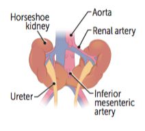
This diagram illustrates the classic renal fusion anomaly where the lower poles are joined by an isthmus. It's usually found at the level of L3-L5 because the IMA blocks its upward migration.
To wrap this up, would you like to try a quick quiz on these anatomical variations, or maybe some flashcards to help you memorize the key clinical associations?
Setting up chat...