AI in radiology: applications in medical imaging and diagnosis
AI in radiology is basically like having a super-powered second pair of eyes that never gets tired! It's moving from being a futuristic concept to a daily reality in many diagnostic centers, helping with everything from spotting tiny fractures to prioritizing urgent brain scans.
Let me pull up some relevant lessons that dive deeper into how these algorithms are actually integrated into clinical practice.
📚 AI Radiology Lessons
The impact of AI in radiology can be broken down into three main "superpowers": Detection, Triage, and Radiomics.
- Computer-Aided Detection (CAD): This is the most common use. AI scans images (like mammograms or chest X-rays) to flag suspicious areas—like a tiny lung nodule or a faint breast mass—that a human might miss during a long shift.
- Triage & Workflow: Imagine a radiologist has 100 CT scans to read. AI can scan them all in seconds and move the one showing an ==Intracranial Hemorrhage (ICH)== to the very top of the list. This "triage" saves lives by reducing time-to-treatment.
- Radiomics: This is where AI extracts data from images that the human eye literally cannot see (like pixel-level texture patterns) to predict things like tumor genetics or response to chemotherapy.
I've put together a quick comparison table to show how AI is shifting the traditional workflow.
| Feature | Traditional Radiology | AI-Assisted Radiology |
|---|---|---|
| Detection | Relies on human visual perception | Automated flagging of abnormalities (CAD) |
| Speed | Limited by human reading time | Near-instantaneous initial screening |
| Consistency | Subject to fatigue and bias | Consistent performance 24/7 |
| Data Usage | Visual interpretation of anatomy | Extraction of "hidden" quantitative data (Radiomics) |
| Prioritization | First-in, first-out (usually) | Urgent cases (e.g., stroke) moved to top of queue |
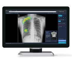
It's one thing to talk about it, but seeing how AI "sees" a scan is pretty cool. It usually highlights findings with bounding boxes or heatmaps. Let me find a visual that shows this in action, like in a chest X-ray or CT scan.
Check out this example of how AI highlights potential issues on a chest X-ray. It uses a heatmap to draw the radiologist's attention to specific areas, like a pulmonary nodule or an apical lesion.
The big takeaway is that AI isn't replacing radiologists—it's augmenting them. The "Radiologist + AI" combo is consistently more accurate than either one alone.
However, we still have to be careful about the "black box" problem (not knowing exactly how the AI reached its conclusion) and ensuring the data used to train these models is diverse. For your exams, keep an eye on how AI is used for automated triage and CAD, as those are the most clinically relevant right now!
Setting up chat...