Abdominal radiology: X-ray, CT and ultrasound interpretation basics
Abdominal imaging is like being a detective—you're looking for clues in different shades of grey to figure out what's happening inside. Whether it's a quick X-ray for a suspected obstruction or a detailed CT for vague pain, each modality has its own "language."
Let me pull up some high-yield lessons that break down these interpretation basics for you.
📚 Abdominal Radiology Lessons
Those lessons are a great starting point for the theory. To help you visualize things, let's look at a classic abdominal X-ray. It's usually the first thing we order when someone comes in with acute "belly pain."
I'll find a clear diagram or image showing the standard landmarks you need to spot.
That X-ray gives us a great "bird's-eye view" of the abdomen. Let's look at how to systematically approach these three main imaging tools.
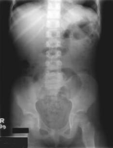
This AP radiograph shows the standard distribution of bowel gas in the stomach and colon. Notice the lumbar spine and pelvic bones, which serve as essential landmarks for orientation.
When you're looking at these images, it's all about knowing what's "normal" so you can spot the "abnormal." Here's a quick comparison of when to use each and what you're looking for.
| Modality | Best For | Key Interpretation Concept |
|---|---|---|
| X-ray (AXR) | Bowel obstruction, perforation (free air), foreign bodies | Gas Patterns: Look for "3-6-9 rule" (Small bowel <3cm, Large bowel <6cm, Cecum <9cm) |
| CT Abdomen | Trauma, malignancy, complex pain, abscesses | Hounsfield Units (HU): Measures density (Water = 0, Air = -1000, Bone = +1000) |
| Ultrasound | Gallstones, biliary tree, appendicitis (kids/lean adults), ascites | Echogenicity: Comparing brightness (Hyperechoic = bright, Hypoechoic = dark) |
That table gives you the "big picture." Now, let's dive into the specifics of how to read each one.
1. Abdominal X-ray (AXR)
The ABDO mnemonic is a lifesaver for a systematic review:
- A - Air: Look for "free air" under the diaphragm (pneumoperitoneum) or abnormal bowel gas patterns.
- B - Bowel: Check the diameter. Remember the
3-6-9 rule: Small bowel should be<3cm, Large bowel<6cm, and the Cecum<9cm. - D - Densities: Look for calcifications (gallstones, kidney stones, or vascular calcification) and foreign bodies.
- O - Organs & Bones: Check the liver/spleen shadows and look for fractures or metastases in the lumbar spine and pelvis.
2. Abdominal CT
CT is the "gold standard" for most acute abdominal pain. It uses Hounsfield Units (HU) to measure density:
- Air (-1000 HU): Appears black.
- Fat (-50 to -100 HU): Dark grey.
- Water (0 HU): Mid-grey.
- Soft Tissue (+40 to +80 HU): Lighter grey.
- Bone/Contrast (+400 to +1000 HU): Bright white.
Pro Tip: Always look for "fat stranding" (hazy, white appearance in the dark grey fat). This is a huge clue that there's inflammation nearby (like in appendicitis or diverticulitis).
3. Abdominal Ultrasound (USG)
Ultrasound is all about echogenicity—how much sound reflects back to the probe:
- Anechoic (Black): Fluid-filled structures like the gallbladder or bladder.
- Hypoechoic (Darker than surroundings): Often seen in solid organs or masses.
- Hyperechoic (Brighter/Whiter): Fat, stones, or gas.
It's the first-line for the biliary tree because it's excellent at spotting gallstones and measuring the common bile duct (CBD).
Would you like to try a quick case study to see how these apply in a real scenario? Or maybe some flashcards to lock in these "3-6-9" and "HU" values?
Setting up chat...