
Trauma/Emergencies — MCQs

On this page
A 78-year-old woman is brought to the emergency department after she fell while gardening and experienced severe pain in her right arm. She has a history of well controlled hypertension and has been found to have osteoporosis. On presentation she is found to have a closed midshaft humerus fracture. No other major findings are discovered on a trauma survey. She is placed in a coaptation splint. The complication that is most associated with this injury has which of the following presentations?
A 56-year-old man presents to the emergency room after being in a motor vehicle accident. He was driving on an icy road when his car swerved off the road and ran head on into a tree. He complains of severe pain in his right lower extremity. He denies loss of consciousness during the accident. His past medical history is notable for poorly controlled hypertension, hyperlipidemia, and major depressive disorder. He takes enalapril, atorvastatin, and sertraline. His temperature is 99.1°F (37.3°C), blood pressure is 155/85 mmHg, pulse is 110/min, and respirations are 20/min. On exam, he is alert and fully oriented. He is unable to move his right leg due to pain. Sensation is intact to light touch in the sural, saphenous, tibial, deep peroneal, and superficial peroneal distributions. His leg appears adducted, flexed, and internally rotated. An anteroposterior radiograph of his pelvis would most likely demonstrate which of the following findings?
A 34-year-old Ethiopian woman who recently moved to the United States presents for evaluation to a surgical outpatient clinic with painful ulceration in her right breast for the last 2 months. She is worried because the ulcer is increasing in size. On further questioning, she says that she also has a discharge from her right nipple. She had her 2nd child 4 months ago and was breastfeeding the baby until the pain started getting worse in the past few weeks, and is now unbearable. According to her health records from Africa, her physician prescribed antimicrobials multiple times with a diagnosis of mastitis, but she did not improve significantly. Her mother and aunt died of breast cancer at 60 and 58 years of age, respectively. On examination, the right breast is enlarged and firm, with thickened skin, diffuse erythema, edema, and an ulcer measuring 3 × 3 cm. White-gray nipple discharge is present. The breast is tender with axillary and cervical adenopathy. Mammography is ordered, which shows a mass with a large area of calcifications, parenchymal distortion, and extensive soft tissue and trabecular thickening in the affected breast. The patient subsequently undergoes core-needle and full-thickness skin punch biospies. The pathology report states a clear dermal lymphatic invasion by tumor cells. Which of the following is the most likely diagnosis?
A 33-year-old woman is brought to the emergency department 15 minutes after being stabbed in the chest with a screwdriver. Her pulse is 110/min, respirations are 22/min, and blood pressure is 90/65 mm Hg. Examination shows a 5-cm deep stab wound at the upper border of the 8th rib in the left midaxillary line. Which of the following structures is most likely to be injured in this patient?
A 65-year-old woman presents to her primary care provider for shoulder pain. She reports that she initially thought the pain was due to "sleeping funny" on the arm, but that the pain has now lasted for 4 weeks. She denies trauma to the joint and says that the pain is worse when reaching overhead to retrieve things from her kitchen cabinets. On physical exam, the patient's shoulders are symmetric, and the right lateral shoulder is tender to palpation. The shoulder has full passive and active range of motion, although pain is reproduced on active abduction of the right arm above 90 degrees. Pain is also reproduced on passively internally rotating and then lifting the shoulder. The patient is able to resist elbow flexion without pain, and she otherwise has 5/5 strength. Which of the following is the most likely diagnosis?
A 62-year-old man comes to the physician for the evaluation of nocturia and a weak urinary stream. These symptoms began 1 year ago, but have progressively worsened over the past 6 months. He now wakes up 3–5 times every night to urinate. He has hypertension treated with hydrochlorothiazide and lisinopril. The patient has smoked a half-pack of cigarettes daily for the past 30 years. He appears well. His temperature is 37.3°C (99.1°F), pulse is 77/min, and blood pressure is 128/77 mm Hg. Cardiopulmonary examination shows no abnormalities. His abdomen is soft and nontender. Digital rectal examination shows a diffusely enlarged prostate with a firm nodule in the right posterior lobe. Urinalysis is within normal limits. Prostate-specific antigen (PSA) level is 6.5 ng/mL (N = 0–4). Which of the following is the most appropriate next step in management?
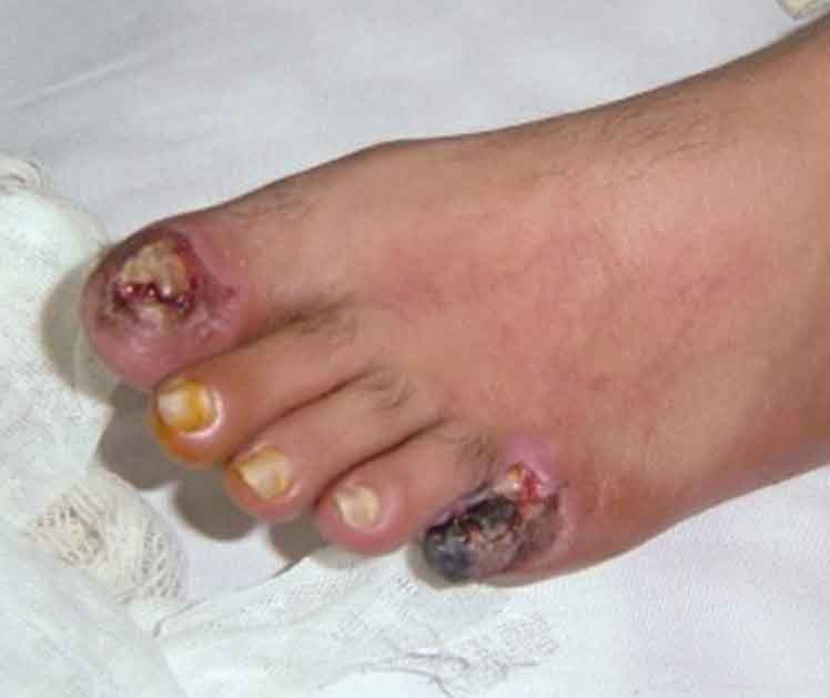
A 75-year-old man presents to the emergency department because of pain in his left thigh and left calf for the past 3 months. The pain occurs at rest, worsens with walking, and is slightly improved by hanging his foot off the bed. He has had hypertension for 25 years and type 2 diabetes mellitus for 30 years. He has smoked 30–40 cigarettes per day for the past 45 years. On examination, the femoral, popliteal, and dorsalis pedis pulses are diminished, but detectable on both sides. The patient’s foot is shown in the image. Which of the following is the most likely diagnosis?
A 67-year-old man comes to the physician for a follow-up examination. He has had lower back pain for several months. The pain radiates down the right leg to the foot. He has no history of any serious illness and takes no medications. His pain increases after activity. The straight leg test is positive on the right. The results of the laboratory studies show: Laboratory test Hemoglobin 14 g/d Leukocyte count 5,500/mm3 with a normal differential Platelet count 350,000/mm3 Serum Calcium 9.0 mg/dL Albumin 3.8 g/dL Urea nitrogen 14 mg/dL Creatinine 0.9 mg/dL Serum immunoelectrophoresis shows an immunoglobulin G (IgG) type monoclonal component of 40 g/L. Bone marrow plasma cells return at 20%. Skeletal survey shows no bone lesions. Magnetic resonance imaging (MRI) shows a herniated disc at the L5. Which of the following is the most appropriate next step?
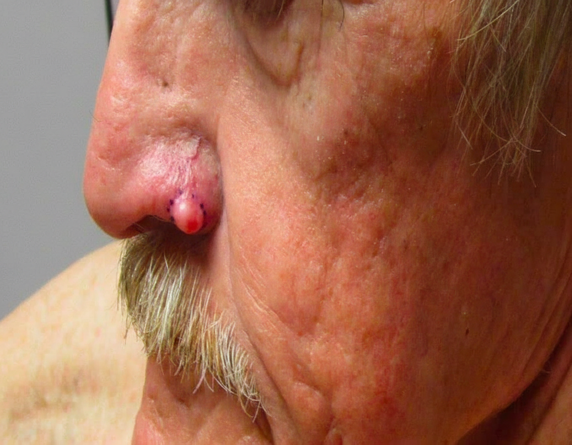
A 67-year-old man is referred to a dermatologist after a reddish mole appears on his nose. The mole’s size has changed over the last 2 years, and occasional bleeding is noted. The man’s medical history is unremarkable, and he does not take any medications. He retired from his construction job 15 years ago. Physical examination of his nose reveals a 2-cm pink papule with a pearly appearance and overlying telangiectasia on the ala of the nose (see image). Which of the following would be the best treatment modality if surgery is not an option?
A 57-year-old woman presents to an outpatient clinic with lower extremity weakness and lower back pain. The patient says that her symptoms began 2 weeks ago when she was working in her garden and have progressively worsened to the extent she currently is unable to walk on her own. She describes the pain as sharp, severe and descending bilaterally from her lower back to her lateral ankles along the posterior surface of her thighs and legs. She also states that she has had several episodes of urinary incontinence for the past couple of days. The patient denies having any similar pain or incontinence in the past. No other significant past medical history. Current medications are alendronate 5 mg orally daily and a daily multivitamin. Her temperature is 37.0℃ (98.6℉), the blood pressure is 110/70 mm Hg, the pulse is 72/min, the respiratory rate is 15/min, and oxygen saturation is 99% on room air. On physical examination, the patient appears to be in significant distress. Strength is ⅗ in her thighs bilaterally and ⅖ in the legs bilaterally left greater than right. Muscle tone is decreased in the lower extremities. The patellar reflex is 1+ bilaterally and plantar reflex is 0+ bilaterally. Fine touch and pain and temperature sensation are decreased in the lower extremities bilaterally, left greater than right. Saddle anesthesia is present. Which of the following is the next, best step in the management of this patient?
Practice by Chapter
Chest trauma management
Practice Questions
Head trauma management
Practice Questions
Spinal trauma
Practice Questions
Blunt abdominal trauma
Practice Questions
Penetrating abdominal trauma
Practice Questions
Pelvic fractures and hemorrhage
Practice Questions
Extremity trauma and vascular injuries
Practice Questions
Burns assessment and management
Practice Questions
Traumatic shock management
Practice Questions
Resuscitative thoracotomy
Practice Questions
Focused Assessment with Sonography in Trauma (FAST)
Practice Questions
Trauma in pregnancy
Practice Questions
Pediatric trauma considerations
Practice Questions
Want unlimited practice?
Get full access to all questions, explanations, and performance tracking.
Scan to download app