
Trauma/Emergencies — MCQs

On this page
A 7-year-old child is brought to the emergency room by his parents in severe pain. They state that he fell on his outstretched right arm while playing with his friends. He is unable to move his right arm which is being supported by his left. On exam, his vitals are normal. His right extremity reveals normal pulses without swelling in any compartments, but there is crepitus above the elbow upon movement. The child is able to flex and extend his wrist, but this is limited by pain. The child has decreased sensation along his thumb and is unable to make the "OK" sign with his thumb and index finger. What is the most likely diagnosis?
A 56-year-old man presents to the emergency department with severe chest pain and a burning sensation. He accidentally drank a cup of fluid at his construction site 2 hours ago. The liquid was later found to contain lye. On physical examination, his blood pressure is 100/57 mm Hg, respiratory rate is 21/min, pulse is 84/min, and temperature is 37.7°C (99.9°F). The patient is sent immediately to the radiology department. The CT scan shows air in the mediastinum, and a contrast swallow study confirms the likely diagnosis. Which of the following is the best next step in the management of this patient’s condition?
A 26-year-old woman comes to the physician because of increasing pain and swelling in her right foot for the past 2 weeks. Initially, the pain was intermittent but it is now constant and she describes it as 8 out of 10 in intensity. She has not had any trauma to the foot or any previous problems with her joints. The pain has not allowed her to continue training for an upcoming marathon. Her only medication is an oral contraceptive. She is a model and has to regularly wear stilettos for fashion shows. She appears healthy. Vital signs are within normal limits. Examination shows swelling of the right forefoot. There is tenderness to palpation over the fifth metatarsal shaft. Pushing the fifth toe inwards produces pain. The remainder of the examination shows no abnormalities. Which of the following is the most likely diagnosis?
A 40-year-old man presents with a painless firm mass in the right breast. Examination shows retraction of the nipple and the skin is fixed to the underlying mass. The axillary nodes are palpable. Which of the following statements is FALSE regarding the above condition?
A 34-year-old man presents to the emergency department by ambulance after being involved in a fight. On arrival, there is obvious trauma to his face and neck, and his mouth is full of blood. Seconds after suctioning the blood, his mouth rapidly fills up with blood again. As a result, he is unable to speak to you. An attempt at direct laryngoscopy fails as a result of his injuries. His vital signs are pulse 102/min, blood pressure 110/75 mmHg, and O2 saturation 97%. Which of the following is indicated at this time?
A 25-year-old man comes to the physician because of right wrist pain after a fall from a ladder. Physical examination shows decreased grip strength and tenderness between the tendons of extensor pollicis longus and extensor pollicis brevis. X-ray of the right wrist shows no abnormalities. This patient is at increased risk for which of the following complications?
A 17-year-old male presents to your office with right knee pain. He is the quarterback of his high school football team and developed the knee pain after being tackled in last night's game. He states he was running with the ball and was hit on the lateral aspect of his right knee while his right foot was planted. Now, he is tender to palpation over the medial knee and unable to bear full weight on the right lower extremity. A joint effusion is present and arthrocentesis yields 50 cc's of clear fluid. Which of the following exam maneuvers is most likely to demonstrate ligamentous laxity?
A previously healthy 5-year-old boy is brought to the emergency department 15 minutes after sustaining an injury to his right hand. His mother says that she was cleaning the bathroom when he accidentally knocked over the drain cleaner bottle and spilled the liquid onto his hand. On arrival, he is crying and holding his right hand in a flexed position. His temperature is 37.7°C (99.8°F), pulse is 105/min, respirations are 25/min, and blood pressure is 105/65 mm Hg. Examination of the right hand shows a 4 x 4 cm area of reddened, blistered skin. The area is very tender to light touch. His ability to flex and extend the right hand are diminished. Radial pulses are palpable. Capillary refill time is less than 3 seconds. Which of the following is the most appropriate next step in management?
A 47-year-old man is brought to the emergency room by his wife. She states that they were having dinner at a restaurant when the patient suddenly became out of breath. His past medical history is irrelevant but has a 20-year pack smoking history. On evaluation, the patient is alert and verbally responsive but in moderate respiratory distress. His temperature is 37°C (98.6°F), blood pressure is 85/56 mm Hg, pulse is 102/min, and respirations are 20/min. His oxygen saturation is 88% on 2L nasal cannula. An oropharyngeal examination is unremarkable. The trachea is deviated to the left. Cardiopulmonary examination reveals decreased breath sounds on the right lower lung field with nondistended neck veins. Which of the following is the next best step in the management of this patient?
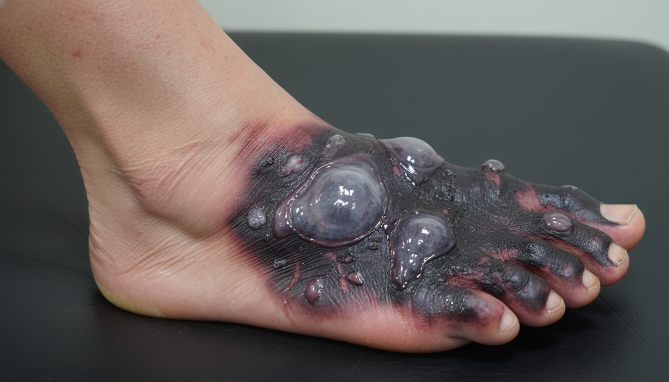
A 60-year-old man presents with pain, swelling, and a purulent discharge from his left foot. He says that the symptoms began 7 days ago with mild pain and swelling on the medial side of his left foot, but have progressively worsened. He states that there has been a foul-smelling discharge for the past 2 days. The medical history is significant for type 2 diabetes mellitus that was diagnosed 10 years ago and is poorly managed, and refractory peripheral artery disease that failed revascularization 6 months ago. The current medications include aspirin (81 mg orally daily) and metformin (500 mg orally twice daily). He has a 20-pack-year smoking history but quit 6 months ago. The family history is significant for type 2 diabetes mellitus in both parents and his father died of a myocardial infarction at 50 years of age. His temperature is 38.9°C (102°F); blood pressure 90/65 mm Hg; pulse 102/min; respiratory rate 22/min; and oxygen saturation 99% on room air. On physical examination, he appears ill and diaphoretic. The skin is flushed and moist. There is 2+ pitting edema of the left foot with blistering and black discoloration (see picture). The lower legs are hairless and the lower extremity peripheral pulses are 1+ bilaterally. Laboratory tests are pending. Blood cultures are positive for Staphylococcus aureus. Which of the following findings is the strongest indication for amputation of the left lower extremity in this patient?
Practice by Chapter
Chest trauma management
Practice Questions
Head trauma management
Practice Questions
Spinal trauma
Practice Questions
Blunt abdominal trauma
Practice Questions
Penetrating abdominal trauma
Practice Questions
Pelvic fractures and hemorrhage
Practice Questions
Extremity trauma and vascular injuries
Practice Questions
Burns assessment and management
Practice Questions
Traumatic shock management
Practice Questions
Resuscitative thoracotomy
Practice Questions
Focused Assessment with Sonography in Trauma (FAST)
Practice Questions
Trauma in pregnancy
Practice Questions
Pediatric trauma considerations
Practice Questions
Want unlimited practice?
Get full access to all questions, explanations, and performance tracking.
Scan to download app