
Trauma/Emergencies — MCQs

On this page
A 24-year-old woman is brought to the emergency department after being assaulted. The paramedics report that the patient was found conscious and reported being kicked many times in the torso. She is alert and able to respond to questions. She denies any head trauma. She has a past medical history of endometriosis and a tubo-ovarian abscess that was removed surgically two years ago. Her only home medication is oral contraceptive pills. Her temperature is 98.5°F (36.9°C), blood pressure is 82/51 mmHg, pulse is 136/min, respirations are 24/min, and SpO2 is 94%. She has superficial lacerations to the face and severe bruising over her chest and abdomen. Her lungs are clear to auscultation bilaterally and her abdomen is soft, distended, and diffusely tender to palpation. Her skin is cool and clammy. Her FAST exam reveals fluid in the perisplenic space. Which of the following is the next best step in management?
A 27-year-old man presents to the emergency department with severe dyspnea and sharp chest pain that suddenly started an hour ago after he finished exercising. He has a history of asthma as a child, and he achieves good control of his acute attacks with Ventolin. On examination, his right lung field is hyperresonant along with diminished lung sounds. Chest wall motion during respiration is asymmetrical. His blood pressure is 105/67 mm Hg, respirations are 22/min, pulse is 78/min, and temperature is 36.7°C (98.0°F). The patient is supported with oxygen, given corticosteroids, and has had analgesic medications via a nebulizer. Considering the likely condition affecting this patient, what is the best step in management?
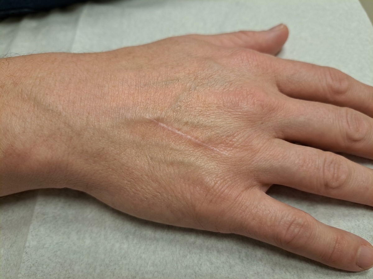
A 16-year-old boy presents to the emergency department after a skateboarding accident. He fell on a broken bottle and received a 4 cm wound on the dorsal aspect of his left hand. His vitals are stable and he was evaluated by the surgeon on call who determined that suturing was not required. After several weeks the wound has almost completely healed (see image). Which of the following is the correct description of this patient's wound before healing?
A 24-year-old man is brought to the emergency department after being involved in a motor vehicle accident as an unrestrained driver. He was initially found unconscious at the scene but, after a few minutes, he regained consciousness. He says he is having difficulty breathing and has right-sided pleuritic chest pain. A primary trauma survey reveals multiple bruises and lacerations on the anterior chest wall. His temperature is 36.8°C (98.2°F), blood pressure is 100/60 mm Hg, pulse is 110/min, and respiratory rate is 28/min. Physical examination reveals a penetrating injury just below the right nipple. Cardiac examination is significant for jugular venous distention. There is also an absence of breath sounds on the right with hyperresonance to percussion. A bedside chest radiograph reveals evidence of a collapsed right lung with depression of the right hemidiaphragm and tracheal deviation to the left. Which of the following is the most appropriate next step in the management of this patient?
A 72-year-old man is brought to the physician for the evaluation of severe nosebleeds and two episodes of bloody vomit over the past 40 minutes. He reports that he has had recurrent nosebleeds almost daily for the last 3 weeks. The nosebleeds last between 30 and 40 minutes. He appears pale. His temperature is 36.5°C (97.7°F), pulse is 95/min, and blood pressure is 110/70 mm Hg. Examination of the nose with a speculum does not show an anterior bleeding source. The upper body of this patient is elevated and his head is bent forward. Cold packs are applied and the nose is pinched at the nostrils for 5–10 minutes. Topical phenylephrine is administered. Despite all measures, the nosebleed continues. Anterior and posterior nasal packing is placed, but bleeding persists. Which of the following is the most appropriate next step in management?
A 58-year-old man with type 2 diabetes mellitus comes to the emergency department because of a 2-day history of dysphagia and swelling in the neck and lower jaw. He has had tooth pain on the left side over the past week, which has made it difficult for him to sleep. Four weeks ago, he had a 3-day episode of flu-like symptoms, including sore throat, that resolved without treatment. He has a history of hypertension. Current medications include metformin and lisinopril. He appears distressed. He is 180 cm (5 ft 11 in) tall and weighs 100 kg (220 lbs); his BMI is 31.6 kg/m2. His temperature is 38.4°C (101.1°F), pulse is 90/min, and blood pressure is 110/80 mm Hg. Oral cavity examination shows a decayed lower left third molar with drainage of pus. There is submandibular and anterior neck tenderness and swelling. His leukocyte count is 15,600/mm3, platelet count is 300,000/mm3, and fingerstick blood glucose concentration is 250 mg/dL. Which of the following is the most likely diagnosis?
A 19-year-old man is brought to the emergency department 35 minutes after being involved in a high-speed motor vehicle collision. On arrival, he is alert, has mild chest pain, and minimal shortness of breath. He has one episode of vomiting in the hospital. His temperature is 37.3°C (99.1°F), pulse is 108/min, respirations are 23/min, and blood pressure is 90/70 mm Hg. Pulse oximetry on room air shows an oxygen saturation of 92%. Examination shows multiple abrasions over his trunk and right upper extremity. There are coarse breath sounds over the right lung base. Cardiac examination shows no murmurs, rubs, or gallop. Infusion of 0.9% saline is begun. He subsequently develops increasing shortness of breath. Arterial blood gas analysis on 60% oxygen shows: pH 7.36 pCO2 39 mm Hg pO2 68 mm Hg HCO3- 18 mEq/L O2 saturation 81% An x-ray of the chest shows patchy, irregular infiltrates over the right lung fields. Which of the following is the most likely diagnosis?
A 68-year-old man comes to the physician because of a 6-month history of difficulty swallowing pieces of meat and choking frequently during meal times. He also sometimes regurgitates foul-smelling, undigested food particles. Examination shows a 3 x 3 cm soft cystic, immobile mass in the upper third of the left side of his neck anterior to the left sternocleidomastoid muscle that becomes prominent when he coughs. A barium swallow shows an accumulation of contrast on the lateral aspect of the neck at the C5 level. Which of the following is the most likely underlying cause for this patient's condition?
A 79-year-old man is admitted to the intensive care unit for hospital acquired pneumonia, a COPD flare, and acute heart failure requiring intubation and mechanical ventilation. On his first night in the intensive care unit, his temperature is 99.7°F (37.6°C), blood pressure is 107/58 mm Hg, and pulse is 150/min which is a sudden change from his previous vitals. Physical exam is notable for jugular venous distension and a rapid heart rate. The ventilator is checked and is functioning normally. Which of the following is the best next step in management for the most likely diagnosis?
An 18-year-old man presents to a rural emergency department after being stabbed multiple times. The patient's past medical history is notable for obesity, diabetes, chronic upper respiratory infections, a 10 pack-year smoking history, and heart failure. He is protecting his airway and he is oxygenating and ventilating well. His temperature is 97.6°F (36.4°C), blood pressure is 74/34 mmHg, pulse is 180/min, respirations are 24/min, and oxygen saturation is 98% on room air. The patient is started on whole blood and the surgeon on call is contacted to take the patient to the operating room. During the secondary survey, the patient complains of shortness of breath. His blood pressure is 54/14 mmHg, pulse is 200/min, respirations are 24/min, and oxygen saturation is 90% on room air. Physical exam is notable for bilateral wheezing on lung exam. The patient goes into cardiac arrest and after 30 minutes, attempts at resuscitation are terminated. Which of the following is associated with this patient's decompensation during resuscitation?
Practice by Chapter
Chest trauma management
Practice Questions
Head trauma management
Practice Questions
Spinal trauma
Practice Questions
Blunt abdominal trauma
Practice Questions
Penetrating abdominal trauma
Practice Questions
Pelvic fractures and hemorrhage
Practice Questions
Extremity trauma and vascular injuries
Practice Questions
Burns assessment and management
Practice Questions
Traumatic shock management
Practice Questions
Resuscitative thoracotomy
Practice Questions
Focused Assessment with Sonography in Trauma (FAST)
Practice Questions
Trauma in pregnancy
Practice Questions
Pediatric trauma considerations
Practice Questions
Want unlimited practice?
Get full access to all questions, explanations, and performance tracking.
Scan to download app