Three days after being admitted to the hospital because of a fall from the roof of a two-story building, a 27-year-old man is being monitored in the intensive care unit. On arrival, the patient was somnolent and not oriented to person, place, or time. A CT scan of the head showed an epidural hemorrhage that was 45 cm3 in size and a midline shift of 7 mm. Emergency surgery was performed with craniotomy and hematoma evacuation on the day of admission. Perioperatively, a bleeding vessel was identified and ligated. Postoperatively, the patient was transferred to the intensive care unit and placed on a ventilator. His temperature is 37°C (98.6°F), pulse is 67/min, and blood pressure is 117/78 mm Hg. The ventilator is set at a FiO2 of 55%, tidal volume of 520 mL, and positive end-expiratory pressure of 5.0 cm H2O. In addition to intravenous administration of fluids, which of the following is the most appropriate next step in managing this patient's nutrition?
A 65-year-old woman comes to the physician because of progressive weight loss for 3 months. Physical examination shows jaundice and a nontender, palpable gallbladder. A CT scan of the abdomen shows an ill-defined mass in the pancreatic head. She is scheduled for surgery to resect the pancreatic head, distal stomach, duodenum, early jejunum, gallbladder, and common bile duct and anastomose the jejunum to the remaining stomach, pancreas, and bile duct. Following surgery, this patient is at the greatest risk for which of the following?
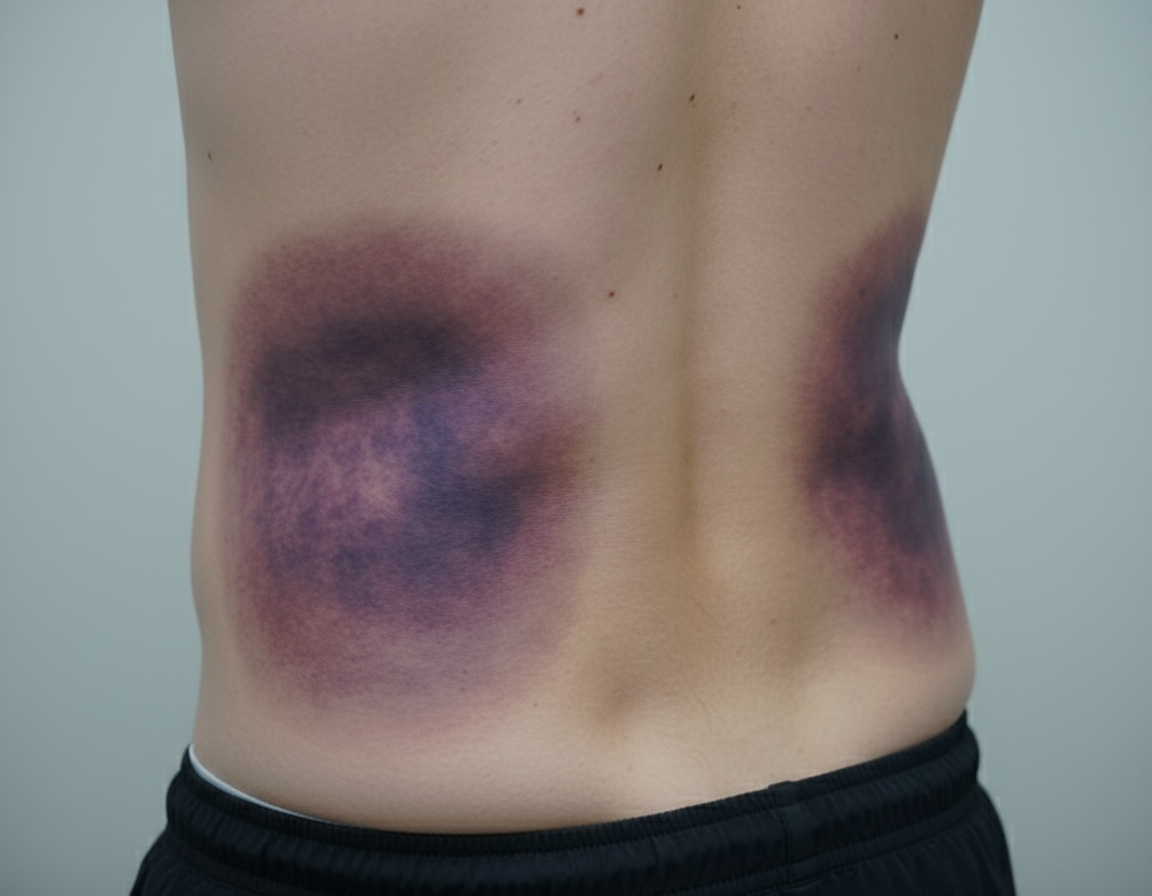
Twelve days after undergoing total pancreatectomy for chronic pancreatitis, a 62-year-old woman notices oozing from her abdominal wound. She first noticed fluid draining 8 hours ago. Her postoperative course has been complicated by persistent hypotension requiring intravenous fluids and decreased ability to tolerate food. She has type 1 diabetes mellitus and glaucoma. The patient smoked one pack of cigarettes daily for 30 years, but quit 2 years ago. She drank a pint of vodka every day starting at age 20 and quit when she was 35 years old. Her current medications include subcutaneous insulin and timolol eye drops. She appears comfortable. Her temperature is 37°C (98.6°F), pulse is 95/min, and blood pressure is 104/78 mm Hg. The abdomen is soft and mildly tender to palpation. There is a 12-cm vertical wound beginning in the epigastrium and extending caudally. 25 mL of a viscous, dark green substance is draining from the middle of the wound. There is a small amount of dried fluid on the patient's hospital gown. The wound edges are nonerythematous. There is no pus draining from the wound. Laboratory studies show: Hematocrit 38% Leukocyte count 8,000/mm3 Serum Na+ 135 mEq/L Cl- 100 mEq/L K+ 3.4 mEq/L HCO3- 23 mEq/L Urea nitrogen 13 mg/dL Creatinine 1.1 mg/dL Glucose 190 mg/dL Which of the following is the most appropriate next step in management?
Three hours after undergoing open proctocolectomy for ulcerative colitis, a 42-year-old male complains of abdominal pain. The pain is localized to the periumbilical and hypogastric regions. A total of 20 mL of urine has drained from his urinary catheter since the end of the procedure. Temperature is 37.2°C (98.9°F), pulse is 92/min, respirations are 12/min, and blood pressure is 110/72 mm Hg. Pulse oximetry on room air shows an oxygen saturation of 99%. Physical examination shows a 20 cm vertical midline incision and an ileostomy in the right lower quadrant. There is no fluid drainage from the surgical wounds. The urinary catheter flushes easily and is without obstruction. Cardiopulmonary examination shows no abnormalities. Serum studies show a blood urea nitrogen of 30 mg/dL and a creatinine of 1.3 mg/dL. Which of the following is the most appropriate next step in management?
One and a half hours after undergoing an elective cardiac catheterization, a 53-year-old woman has right flank and back pain. She has hypertension, hypercholesterolemia, and type 2 diabetes mellitus. She had an 80% stenosis in the left anterior descending artery and 2 stents were placed. Intravenous unfractionated heparin was used prior to the procedure. Prior to admission, her medications were enalapril, simvastatin, and metformin. Her temperature is 37.3°C (99.1°F), pulse is 102/min, and blood pressure is 109/75 mm Hg. Examination shows a tender lower abdomen; there is no guarding or rigidity. There is right suprainguinal fullness and tenderness. There is no bleeding or discharge from the femoral access site. Cardiac examination shows no murmurs, rubs, or gallops. Femoral and pedal pulses are palpable bilaterally. 0.9% saline infusion is begun. A complete blood count shows a hematocrit of 36%, leukocyte count of 8,400/mm3, and a platelet count of 230,000/mm3. Which of the following is the most appropriate next step in management?
A 45-year-old man undergoes elective vasectomy for permanent contraception. The procedure is performed under local anesthesia. There are no intra-operative complications and he is discharged home with ibuprofen for post-operative pain. This patient is at increased risk for which of the following complications?
Ten days after undergoing emergent colectomy for a ruptured bowel that she sustained in a motor vehicle accident, a 59-year-old woman has abdominal pain. During the procedure, she was transfused 3 units of packed red blood cells. She is currently receiving total parenteral nutrition. Her temperature is 38.9°C (102.0°F), pulse is 115/min, and blood pressure is 100/60 mm Hg. Examination shows tenderness to palpation in the right upper quadrant of the abdomen. Bowel sounds are hypoactive. Serum studies show: Aspartate aminotransferase 142 U/L Alanine aminotransferase 86 U/L Alkaline phosphatase 153 U/L Total bilirubin 1.5 mg/dL Direct bilirubin 1.0 mg/dL Amylase 20 U/L Which of the following is the most likely diagnosis?
A 28-year-old man comes to the physician because of increasing shortness of breath, abdominal fullness, and pedal edema for 3 months. Four months ago, he was diagnosed with pulmonary tuberculosis and is currently receiving therapy with isoniazid, rifampin, pyrazinamide, and ethambutol. His temperature is 37°C (98.6°F), pulse is 100/min, respirations are 20/min and blood pressure is 96/70 mm Hg. Examination shows 2+ pretibial edema bilaterally. There is jugular venous distention. The jugular venous pressure rises with inspiration. Breath sounds are decreased at lung base bilaterally. Cardiac examination reveals an early diastolic sound over the left sternal border. The abdomen is distended and shifting dullness test is positive. An ECG shows low-amplitude QRS complexes. Chest x-ray shows small pleural effusions bilaterally and calcifications over the left cardiac silhouette. Echocardiography shows a 40% decrease in the velocity of peak diastolic blood flow across the mitral valve during inspiration. A cardiac catheterization shows elevated right ventricular diastolic pressure with characteristic dip-and-plateau waveform. Which of the following is the most appropriate next step in management?
A 55-year-old woman is being managed on the surgical floor after having a total abdominal hysterectomy as a definitive treatment for endometriosis. On day 1 after the operation, the patient complains of fevers. She has no other complaints other than aches and pains from lying in bed as she has not moved since the procedure. She is currently receiving ondansetron, acetaminophen, and morphine. Her temperature is 101°F (38.3°C), blood pressure is 127/68 mmHg, pulse is 100/min, respirations are 17/min, and oxygen saturation is 94% on room air. Her abdominal exam is within normal limits and cardiopulmonary exam is only notable for mild crackles. Which of the following is the most likely etiology of this patient’s fever?
A 56-year-old woman comes to the physician because she palpated a mass in her right breast during self-examination a week ago. Menarche was at the age of 14, and her last menstrual period was at the age of 51. Vital signs are within normal limits. Examination shows a nontender, firm and hard mass in the upper outer quadrant of the right breast. Mammography shows large, dense breasts, with a 1.7-cm mass in the right upper outer quadrant. The patient undergoes right upper outer quadrant lumpectomy with subsequent sentinel node biopsy, which reveals moderately differentiated invasive ductal carcinoma and micrometastasis to one axillary lymph node. There is no evidence of extranodal metastasis. The tumor tests positive for both estrogen and progesterone receptors and does not show human epidermal growth factor receptor 2 (HER2) over-expression. Flow-cytometry reveals aneuploid tumor cells. Which of the following factors has the greatest effect on this patient's prognosis?
Explanation: ***Enteral feeding via nasogastric tube*** - This patient has been **somnolent** and on a ventilator for 3 days after a significant head injury, indicating a prolonged period without oral intake and an inability to protect his airway for oral feeding. **Early enteral nutrition** via a nasogastric tube is preferred in critically ill patients, especially those with head injuries, as it helps maintain gut integrity and reduces complications compared to parenteral nutrition. - The patient's **hemodynamic stability** (blood pressure and pulse are within a reasonable range for a ventilated patient) suggests he can tolerate enteral feeding, and there are no signs of gut ischemia or ileus that would contraindicate it. *Enteral feeding using a percutaneous endoscopic gastrostomy (PEG) tube* - While a PEG tube provides enteral nutrition, it is typically reserved for patients requiring **long-term enteral support** (usually more than 4-6 weeks) or those who cannot tolerate a nasogastric tube. - Given that it has only been 3 days post-injury, a **less invasive method** like a nasogastric tube is initially preferred. *Oral feeding* - The patient is described as **somnolent** and on a ventilator, meaning he is not awake enough or able to protect his airway to safely receive oral feeding. - Attempting oral feeding in this state carries a high risk of **aspiration pneumonia**. *Keep patient NPO* - Keeping the patient NPO (nil per os) for an extended period in critical illness is associated with several negative outcomes, including **gut mucosal atrophy, increased infection risk**, and poorer clinical outcomes. - After 3 days, initiating nutritional support is crucial to prevent these complications. *Total parenteral nutrition* - **Total parenteral nutrition (TPN)** is generally considered a last resort when the gastrointestinal tract is non-functional or enteral feeding is contraindicated. - TPN is associated with a **higher risk of complications**, such as central line infections, liver dysfunction, and metabolic disturbances, compared to enteral feeding.
Explanation: ***Microcytic anemia*** - The surgical procedure described is a **Whipple procedure**, which involves partial gastrectomy and duodenectomy. This significant alteration to the upper GI tract can lead to **iron malabsorption**, as iron is primarily absorbed in the duodenum and proximal jejunum, and gastric acid is crucial for converting dietary iron to its absorbable ferrous form. - **Iron deficiency** is the most common cause of **microcytic anemia**, characterized by small, pale red blood cells, due to impaired hemoglobin synthesis as a result of insufficient iron availability for the heme component. *Wide-based gait* - A **wide-based gait** is typically associated with **ataxia** or conditions affecting cerebellar function or proprioception, which are not direct complications of a Whipple procedure. - While nutritional deficiencies can occur post-surgery, a wide-based gait specifically points to neurological impairment rather than postsurgical metabolic issues. *Calcium oxalate kidney stones* - **Calcium oxalate kidney stones** are often associated with conditions causing **hypercalciuria** or malabsorption of fat, which leads to increased oxalate absorption in the colon. While fat malabsorption can occur after a Whipple due to pancreatic insufficiency, dietary oxalate intake and hydration status are generally more significant determinants of stone formation. - The surgery itself does not directly increase the risk for calcium oxalate kidney stones more than other listed complications. *Increased bile production* - A Whipple procedure involves the removal of the **gallbladder** and rerouting of the **bile duct** directly into the jejunum. This does not lead to increased bile production, but rather a different regulation and flow of bile. - In fact, the absence of the gallbladder means there is no storage for bile, leading to a continuous, unregulated flow of bile into the small intestine, potentially contributing to maldigestion or diarrhea, but not "increased production." *Hypercoagulable state* - While surgery, including a Whipple procedure, can transiently increase the risk of a **hypercoagulable state** (e.g., deep vein thrombosis, pulmonary embolism) in the immediate postoperative period due to immobility and tissue injury, this risk is generally mitigated with prophylactic anticoagulation. - The question asks about the **greatest risk** post-surgery, and long-term complications related to altered anatomy and malabsorption, such as microcytic anemia, are more direct and sustained consequences unique to the extent of the resection.
Explanation: ***Total parenteral nutrition and ostomy pouch*** - The patient, having undergone total pancreatectomy, is experiencing an **enterocutaneous fistula** from an anastomotic leak (e.g., gastrojejunostomy or choledochojejunostomy), presenting as viscid, dark green drainage from the wound. - An **ostomy pouch** will protect the skin from digestive enzymes and allow quantification of output, while **total parenteral nutrition (TPN)** provides bowel rest, promoting spontaneous healing of the fistula. - This conservative approach is appropriate given the patient's **hemodynamic stability** and **absence of signs of sepsis or peritonitis**. *Intravenous antibiotic therapy* - While infection is a concern, there are **no clear signs of systemic infection** (e.g., fever, leukocytosis, erythema, purulent drainage) in this patient. - **Antibiotics alone** would not address the underlying anatomical leak and its resulting drainage. *Surgical exploration of the abdomen* - **Surgical exploration** is typically reserved for cases with signs of **sepsis**, **peritonitis**, **uncontrolled leak**, or failure of conservative management. - Given the patient is hemodynamically stable without signs of severe infection or acute abdomen, a **conservative approach** is preferred initially. *Wound debridement and irrigation* - **Wound care** is important, but debridement and irrigation alone will **not stop the internal leakage** of enteric content from the gastrointestinal tract. - The primary issue is an internal fistula, not a localized wound infection requiring debridement. *Oral food intake and intravenous fluid administration* - **Oral food intake** would exacerbate the fistula by stimulating digestive secretions and increasing enteric output, hindering natural healing. - While **intravenous fluids** are used for hydration and electrolyte balance, they do not address the fistula, and **bowel rest is crucial** for spontaneous closure.
Explanation: ***Administer intravenous fluids*** - The patient's **oliguria** (20 mL urine over 3 hours, ~7 mL/hour) post-surgery, elevated **BUN (30 mg/dL)**, and **creatinine (1.3 mg/dL)** with a **BUN:Cr ratio >20:1** suggest **prerenal acute kidney injury** due to **hypovolemia**. - Post-operative fluid losses from **third-spacing**, blood loss, and insensible losses commonly cause hypovolemia after major abdominal surgery. - **Intravenous fluids** are the most appropriate initial step to restore intravascular volume and improve renal perfusion. *Administer tamsulosin* - **Tamsulosin** is an alpha-blocker used to relax smooth muscle in the prostate and bladder neck, primarily for **urinary retention** due to benign prostatic hyperplasia. - This patient's oliguria is due to **prerenal azotemia** from hypovolemia, not prostatic obstruction, and the catheter flushes easily without obstruction. *Administer intravenous furosemide* - **Furosemide** is a loop diuretic that increases urine output, but administering it in the context of **prerenal acute kidney injury** can worsen hypovolemia and further compromise renal function. - Diuretics are generally contraindicated in oliguria due to hypovolemia and should only be considered after volume resuscitation. *Obtain an abdominal CT* - While an abdominal CT can diagnose surgical complications, there are no immediate signs of a surgical emergency like **anastomotic leak** or **bowel obstruction**. - Addressing the likely **hypovolemia** is more urgent and should precede further imaging in this scenario. *Start ciprofloxacin* - The patient does not show signs of infection, such as fever or localized signs of bacterial peritonitis, making **antibiotics** like ciprofloxacin inappropriate as the initial management step. - The elevated BUN and creatinine are more indicative of volume depletion than infection.
Explanation: ***CT scan of the abdomen and pelvis*** - The patient's symptoms of **right flank and back pain**, along with **right suprainguinal fullness and tenderness** following cardiac catheterization with **anticoagulation**, are highly suggestive of a **retroperitoneal hemorrhage**. - A **CT scan of the abdomen and pelvis** is the most appropriate diagnostic tool to confirm the presence, size, and location of the retroperitoneal bleed. *X-ray of the abdomen* - An **X-ray of the abdomen** is generally not useful for diagnosing soft tissue conditions like retroperitoneal hemorrhage. - It would be primarily used for detecting **ureteral stones** or **bowel obstruction**, which are not the primary concerns with this presentation. *Obtain an ECG* - While an **ECG** is important in cardiac patients, the current symptoms of flank pain, back pain, and suprainguinal fullness are not typical signs of an acute cardiac event. - The patient just underwent a successful cardiac catheterization and stent placement, making **ischemia** less likely to be the immediate cause of these specific symptoms. *Administer protamine sulfate* - **Protamine sulfate** is used to reverse the effects of **unfractionated heparin**, which was used prior to the procedure. - While a retroperitoneal hemorrhage is suspected, reversal of heparin without definitive diagnosis and assessment of the bleeding severity could be premature and might increase the risk of **thrombosis** in a patient with recent stent placement. *Administer intravenous atropine* - **Intravenous atropine** is used to treat **bradycardia**. - The patient's pulse is 102/min, indicating **tachycardia**, not bradycardia, making atropine an inappropriate treatment.
Explanation: **Sperm granuloma** - A **sperm granuloma** can occur after vasectomy due to the extravasation of sperm from the severed vas deferens, leading to a foreign body granulomatous reaction. - This complication presents as a **palpable, tender nodule** at the vasectomy site and is a relatively common long-term issue. *Prostatitis* - **Prostatitis** is an inflammation of the prostate gland, and there is no direct mechanistic link or increased risk following a vasectomy. - It is typically caused by bacterial infection or non-infectious inflammatory processes, unrelated to the **vas deferens** ligation. *Seminoma* - **Seminoma** is a type of testicular germ cell tumor, and extensive research has shown no increased risk of developing testicular cancer after vasectomy. - The procedure does not alter the cellular processes or environment within the testicles that predispose to germ cell tumor formation. *Testicular torsion* - **Testicular torsion** is a urological emergency involving the twisting of the spermatic cord, which cuts off blood supply to the testis. - This condition is not associated with vasectomy; it typically occurs due to an anatomical abnormality (e.g., **bell-clapper deformity**) or trauma. *Inguinal hernia* - An **inguinal hernia** is a protrusion of abdominal contents through a weakness in the abdominal wall, specifically in the inguinal canal. - Vasectomy is a superficial procedure that does not involve manipulating or weakening the abdominal wall in a way that would increase the risk of an inguinal hernia.
Explanation: ***Acalculous cholecystitis*** - This patient's clinical picture of **fever**, **RUQ tenderness**, **leukocytosis**, and mildly elevated liver enzymes in the setting of recent **major surgery**, **trauma**, and **total parenteral nutrition (TPN)** is highly suggestive of **acalculous cholecystitis**. - **Acalculous cholecystitis** often occurs in critically ill patients due to gallbladder stasis, ischemia, and inflammation, usually without the presence of stones. *Hemolytic transfusion reaction* - While the patient received blood transfusions, a **hemolytic transfusion reaction** typically presents with fever, chills, flank pain, and **hemoglobinuria**, none of which are explicitly mentioned. - Liver enzyme elevations can occur, but the significant RUQ tenderness and absence of signs of hemolysis make it less likely. *Acute cholecystitis (calculous)* - **Acute cholecystitis with gallstones** typically presents with similar symptoms to acalculous cholecystitis (pain, fever), but requires the presence of gallstones causing obstruction. - The clinical context of critical illness, recent surgery, and TPN use points more towards acalculous inflammation rather than stone-related disease. *Small bowel obstruction* - **Small bowel obstruction** would present with more pronounced **abdominal distention**, **vomiting**, and often **high-pitched bowel sounds** followed by absent sounds, which is not the primary picture here. - Although bowel sounds are hypoactive, the focal RUQ tenderness and liver enzyme changes are not typical of a primary small bowel obstruction. *Acute pancreatitis* - **Acute pancreatitis** is usually characterized by **severe epigastric pain** radiating to the back, and significantly elevated **amylase** and **lipase** levels. - The patient's amylase is normal, and lipase is not mentioned but usually tracks with amylase in pancreatitis.
Explanation: ***Pericardiectomy*** - The patient's symptoms (shortness of breath, edema, ascites, elevated JVP with Kussmaul sign, early diastolic sound, low voltage ECG, calcified pericardium on CXR, and inspiratory decrease in mitral inflow velocity) are classic for **constrictive pericarditis**, often a complication of **tuberculosis**. - **Pericardiectomy** is the definitive treatment for constrictive pericarditis, involving surgical removal of the thickened, fibrotic pericardium to relieve the heart from external compression. *Colchicine therapy* - **Colchicine** is primarily used in the management of **acute pericarditis** or recurrent pericarditis to reduce inflammation. - It is not effective for **constrictive pericarditis**, where the pathology involves fixed fibrous thickening rather than active inflammation. *Heart transplantation* - **Heart transplantation** is a treatment option for **end-stage heart failure** refractory to other medical or surgical interventions. - While constrictive pericarditis can lead to heart failure symptoms, it is a mechanical issue with the pericardium, and relieving this constriction via pericardiectomy is the primary treatment, not replacing the heart. *Implantable cardioverter defibrillator* - An **ICD** is indicated for patients at high risk of sudden cardiac death due to **ventricular arrhythmias** or severe left ventricular dysfunction. - It does not address the underlying mechanical restriction of **constrictive pericarditis** and would not improve the patient's symptoms related to cardiac filling. *Metoprolol therapy* - **Metoprolol**, a beta-blocker, is used to reduce heart rate, blood pressure, and myocardial oxygen demand in conditions like **heart failure with reduced ejection fraction**, angina, or arrhythmias. - It is generally **contraindicated** or used with extreme caution in **constrictive pericarditis** because further slowing the heart rate can worsen cardiac output in a heart already constrained by a rigid pericardium, and it does not resolve the mechanical constriction.
Explanation: ***Inflammatory stimulus of surgery*** - Postoperative fever occurring within the first 24-48 hours after surgery, especially a major abdominal procedure, is most commonly due to the **systemic inflammatory response** to tissue trauma and stress from the surgery itself. - The temperature of 101°F (38.3°C) is a common reactive fever. In this timeframe, **atelectasis** (part of the inflammatory response to surgery) is the classic cause, supported by the patient's **immobility since surgery** and **mild crackles** on exam. - The patient has no other specific signs of infection, making this the most likely cause. *Deep vein thrombosis* - While DVT is a concern post-surgery, it typically presents with **leg pain, swelling, and tenderness**, not primarily as fever alone on day 1. - A fever from DVT would usually indicate a more advanced complication like pulmonary embolism, which is inconsistent with the mild crackles and stable oxygen saturation. *Abscess formation* - Abscesses usually take several days to form and present with significant fevers, localized pain, and possibly purulent drainage, not typically within the first **24 hours post-op**. - The abdominal exam is noted as within normal limits, making an abscess unlikely at this early stage. *Urinary tract infection* - UTIs are common post-op, especially with catheterization, but typically present with **dysuria, frequency, urgency**, and sometimes suprapubic pain, which are absent here. - While fever can be a symptom, the lack of urinary complaints makes it a less likely primary diagnosis on day 1. *Wound infection* - Wound infections rarely develop within the first **24-48 hours** post-surgery, as bacteria require time to proliferate and cause inflammatory signs. - Typical signs include **erythema, warmth, tenderness, and purulent drainage** at the incision site, which are not mentioned.
Explanation: **Nodal status** - The presence of **micrometastasis to one axillary lymph node** is the *most significant prognostic indicator* in this patient's case. - **Lymph node involvement** signifies systemic spread and is the strongest predictor of recurrence and overall survival in breast cancer. *Age* - While **age** can influence treatment choices and comorbidity, it is generally *less impactful on long-term prognosis* than nodal status. - Very young or very old age can sometimes be associated with more aggressive disease or worse outcomes, but it is not the primary determinant. *Tumor size* - The **tumor size of 1.7 cm** is a prognostic factor, with larger tumors generally having a worse prognosis. - However, for this patient, the **presence of lymph node metastasis** is a more powerful indicator of systemic disease than the primary tumor size alone. *Hormone receptor status* - **Positive estrogen and progesterone receptors** indicate that the tumor is likely to respond to endocrine therapies. - This is a *favorable prognostic factor* as it opens up additional treatment options, but it does not outweigh the negative impact of nodal involvement. *HER2 receptor status* - **Absence of HER2 overexpression** is a positive factor, as HER2-positive cancers are generally more aggressive and require targeted therapy. - However, while HER2 status guides treatment, the presence of **lymph node metastasis** still holds greater weight in determining overall prognosis.
Explanation: ***Vascularized lymph node transfer*** - This patient presents with **secondary lymphedema** due to axillary dissection and radiotherapy, which has not responded to conservative management. - **Vascularized lymph node transfer** is a surgical option that involves transplanting healthy lymph nodes to the affected area to re-establish lymphatic drainage pathways, offering a more definitive solution for refractory cases. *Diethylcarbamazine* - **Diethylcarbamazine** is an anti-filarial drug used to treat lymphedema caused by **parasitic infections**, specifically filariasis. - The patient's lymphedema is secondary to breast cancer treatment, not parasitic infection, making this a **misdirected treatment**. *Low molecular weight heparin* - **Low molecular weight heparin** is an anticoagulant used to prevent or treat **venous thromboembolism (VTE)**. - While patients with cancer are at increased risk for VTE, her symptoms are consistent with lymphedema and not thrombosis, which would typically present with more acute pain and swelling, making this an inappropriate treatment. *Endovascular stenting* - **Endovascular stenting** is a procedure used to open blocked or narrowed **blood vessels**, such as in peripheral artery disease or venous obstruction. - Her condition is specifically lymphedema, a lymphatic circulation issue, not a vascular obstruction, so stenting would not address the underlying problem. *Antibiotics* - **Antibiotics** are used to treat **bacterial infections**, which can complicate lymphedema (e.g., cellulitis). - While chronic lymphedema causes skin changes (hyperkeratosis, papillomatosis, induration), the patient shows no signs of **acute infection** such as fever, pain, erythema, or warmth, making empirical antibiotics unnecessary at this stage.
Explanation: **Splinting** - **Nocturnal wrist splinting** is often the first-line treatment for **carpal tunnel syndrome (CTS)**, especially when symptoms are worse at night, by maintaining the wrist in a neutral position to reduce pressure on the median nerve. - If nocturnal symptoms are severe or persistent during the day, **daytime splinting** may also be beneficial, providing both immediate relief and long-term symptom management. *Nonsteroidal anti-inflammatory drugs* - **NSAIDs** are generally not effective for **carpal tunnel syndrome** as it is a compressive neuropathy, not primarily an inflammatory condition of a joint or muscle. - While NSAIDs can help with general pain, they do not address the underlying **median nerve compression**. *Electromyography testing* - **Electromyography (EMG)** and **nerve conduction studies (NCS)** are diagnostic tests used to confirm the diagnosis and assess the severity of **nerve damage**, but they are not a treatment. - These tests are typically performed if initial conservative management fails or if surgery is being considered, to help guide treatment decisions. *Corticosteroid injection* - **Corticosteroid injections** can provide temporary relief by reducing inflammation and swelling around the **median nerve**. - While effective, they are usually considered after conservative measures like splinting have been tried and failed, or for more severe cases where immediate but temporary symptom relief is desired before other definitive treatments. *Surgical decompression* - **Surgical decompression** (carpal tunnel release) is the most definitive treatment for **carpal tunnel syndrome** and is typically reserved for cases that have failed extensive conservative management. - Given that this is the patient's initial presentation for symptoms, **less invasive methods** should be attempted first.
Explanation: ***Wound infection*** - The patient presents with **fever**, worsening **lower abdominal pain**, **tachycardia**, and **local signs of inflammation** (redness around the surgical site, tenderness) one week post-hysterectomy, with a **leukocytosis and left shift** (elevated neutrophils and bands). These findings are highly characteristic of a common **post-surgical wound infection**. - The lack of significant drainage initially does not rule out infection, and the symptoms are localized to the surgical area. *Surgical error* - The operative report and intraoperative cystoscopy indicated the surgery was **uncomplicated**, making an immediate post-operative surgical error less likely to be the primary cause of these symptoms. - While complications can arise later, the current presentation points more directly to an infectious process rather than an unnoted immediate surgical complication. *Post-operative ileus* - Although bowel sounds are hypoactive, the patient is **tolerating an oral diet well** and denies nausea, vomiting, or abdominal distension, which are key symptoms of a clinically significant ileus. - Her primary complaint is localized pain and systemic signs of infection, rather than generalized abdominal distension and inability to pass flatus or stool. *Alcohol withdrawal* - While the patient has a history of alcohol abuse, the primary symptoms (fever, localized abdominal pain, redness around the incision, leukocytosis) are more indicative of an **infectious process** than alcohol withdrawal. - Alcohol withdrawal typically presents with tremors, agitation, hallucinations, and autonomic instability, and while some overlap (tachycardia) exists, the overall clinical picture doesn't fit. *Urinary tract infection* - The urine analysis shows **negative nitrites and leukocyte esterase**, with only 2 WBC/hpf, which makes a **urinary tract infection (UTI) highly unlikely** despite the presence of some bacteria (50 cfu/mL, which is often considered contamination in a clean catch). - The patient's symptoms are also predominantly localized to the surgical wound area rather than dysuria, frequency, or urgency.
Explanation: ***Insufficient mechanical hemostasis*** - The patient's **coagulation studies are within normal limits** (normal PT, aPTT, bleeding time, and platelet count), ruling out most common intrinsic bleeding disorders. - Given the timing (8 hours post-surgery) and the nature of bleeding (oozing from incision margins and colostomy site), **inadequate surgical closure or ligature** is the most probable cause. *Factor VIII deficiency* - This would present with a **prolonged activated partial thromboplastin time (aPTT)**, which is normal in this patient (36 seconds). His brother's death from a bleeding disorder is a red herring. - Congenital factor deficiencies typically manifest earlier in life and cause more severe, spontaneous bleeding, not just post-operative oozing with normal coagulation factors. *Liver dysfunction* - Severe liver dysfunction would typically lead to **prolonged PT and aPTT** due to impaired synthesis of clotting factors. - While the patient has elevated GGT, indicating some liver stress likely from alcohol, his AST and ALT are only mildly elevated, and his coagulation tests are normal. *Erosion of blood vessels* - This is less likely to cause widespread oozing and would typically present as a more significant, **pulsatile hemorrhage** or hematoma. - While possible in a surgical field, the lack of significant hemodynamic compromise and normal coagulation points away from a major vessel erosion. *Platelet dysfunction* - This would typically result in a **prolonged bleeding time**, which is normal in this patient (4 minutes). - Although the patient was on aspirin, it was stopped 7 days prior to surgery, which is typically sufficient for platelet function to recover.
Explanation: ***Venous insufficiency*** - The presence of **edema**, **dilated tortuous veins**, and **yellow-brown spots** (hemosiderin deposition) on the lower extremities, along with a **painless, shallow, exudative ulcer** above the medial ankle, are classic signs of chronic venous insufficiency. - The ulcer's location (medial malleolus) and its characteristics (granulation tissue, oozing) further support a venous etiology, as good arterial pulses indicate adequate inflow. *Peripheral neuropathy* - Ulcers due to peripheral neuropathy (e.g., in diabetes) are typically **painless** but often occur on the **plantar surface of the foot** or pressure points and can be deep. - While the patient has diabetes, the clinical presentation with prominent venous stasis signs and edema points away from a primary neuropathic ulcer in this location. *Drug-induced microvascular occlusion* - Drug-induced microvascular occlusion (e.g., from **warfarin necrosis**) typically presents as painful, irregular, purpuric lesions that can progress to necrosis and ulceration, often occurring within days of starting the medication or with high doses. - The 3-month history, painless nature, and specific signs of venous stasis do not align with drug-induced microvascular occlusion. *Chronic pressure* - Pressure ulcers develop over **bony prominences** due to prolonged pressure, leading to tissue ischemia and breakdown. - This patient's ulcer is above the medial ankle, which is not a common site for pressure ulcers, and the presentation includes clear signs of venous hypertension, not just external compression. *Decreased arterial blood flow* - Ulcers due to decreased arterial blood flow (arterial ulcers) are typically **painful**, often located on the **toes, heels, or dorsum of the foot**, and have a "punched-out" appearance with **pale bases** and minimal granulation tissue. - The presence of palpable pedal pulses and the painless nature of the ulcer rule out significant arterial insufficiency as the primary cause.
Explanation: ***Physical therapy*** - This patient presents with **shoulder impingement syndrome**, likely due to his occupation as a painter. **Physical therapy** is the first-line treatment, focusing on strengthening rotator cuff muscles, improving posture, and increasing range of motion. - The positive response to a **lidocaine injection** into the subacromial space confirms the diagnosis and indicates that an inflammatory process (like **tendinitis** or **bursitis**) is the source of pain, which usually responds well to conservative management. *Thoracic outlet decompression* - **Thoracic outlet syndrome** is characterized by neurovascular compression, leading to symptoms like **paresthesia, numbness, or weakness in the arm/hand**, which are absent in this patient. - Surgical decompression is a last-resort treatment for severe, refractory cases of thoracic outlet syndrome and is not indicated here. *Arthroscopic repair* - **Arthroscopic repair** is usually reserved for cases of **rotator cuff tears** that fail conservative management or for large, traumatic tears. There is no evidence of a tear in this case, and the pain relief with lidocaine suggests inflammation, not structural damage requiring surgery. - It would be a premature and overly aggressive intervention given the patient's symptoms and initial response to diagnostic injection. *Intraarticular glucocorticoids* - While **glucocorticoid injections** can reduce inflammation and provide temporary pain relief in shoulder impingement, they are typically considered after a trial of conservative management, such as **physical therapy**, has failed. - Repeated injections carry risks like **tendon weakening** and may mask symptoms, delaying appropriate long-term management. *MRI of the shoulder* - An **X-ray was already performed** and showed no abnormalities, ruling out significant bony pathology. An **MRI** would be indicated if there was suspicion of a **rotator cuff tear** or other significant soft tissue injury that has not responded to initial conservative measures. - Given the classic presentation of impingement and the positive response to a diagnostic injection, immediate MRI is not necessary for establishing the diagnosis or guiding the next step in initial treatment.
Explanation: ***Sharp surgical debridement of the ulcer*** - The presence of a **painless ulcer**, decreased sensation (neuropathy), and a calloused margin with loose tissue indicates a **neuropathic ulcer** common in diabetic patients. **Sharp surgical debridement** is crucial to remove non-viable tissue and promote healing. - The probe reaching the deep plantar space suggests a potential deep infection or osteomyelitis, which needs aggressive debridement to remove all infected and necrotic tissue. However, since the X-ray is normal, it is less likely to have osteomyelitis, but it needs to be ruled out by further investigations. *Surgical revascularization of the right foot* - This is primarily indicated for **ischemic ulcers** where blood supply is compromised. - The patient has **intact pedal pulses**, meaning good distal blood flow, making revascularization unnecessary at this stage. *Amputation of the right forefoot* - Amputation is a measure of last resort for **severe, non-healing ulcers** with extensive infection or gangrene that fail to respond to conservative and surgical debridement. - The current presentation does not warrant such an extreme intervention as a first step. *Total contact casting of right foot* - **Total contact casting** is used for **off-loading pressure** from a neuropathic ulcer to facilitate healing. - While it's an important step in management, it should generally follow **debridement** to ensure a clean wound bed. *Intravenous antibiotic therapy* - This is indicated if there are definitive signs of **spreading infection** (e.g., cellulitis, fever, purulence). - While debridement helps prevent infection by removing necrotic tissue, there is no mention of systemic signs of infection or severe local infection requiring immediate IV antibiotics.
Explanation: **Lumpectomy with sentinel lymph node biopsy, followed by radiation and hormone therapy** - The patient has **early-stage (T1N0M0) estrogen receptor (ER)-positive, HER2-negative invasive ductal carcinoma** suitable for **breast-conserving surgery (lumpectomy)**. - **Lumpectomy** must be followed by **radiation therapy** to the remaining breast tissue to reduce the risk of local recurrence, and **endocrine therapy** (due to ER positivity) is indicated to reduce systemic recurrence risk. - **Sentinel lymph node biopsy** is performed to stage the axilla; if positive, an axillary lymph node dissection may be indicated. However, in this case, the mass is small, and there is no palpable axillary adenopathy, making sentinel lymph node biopsy the appropriate initial step. *Lumpectomy with sentinel lymph node biopsy followed by hormone therapy* - While **lumpectomy with sentinel lymph node biopsy** and **hormone therapy** are part of the appropriate management, **radiation therapy** to the conserved breast is a critical component that is missing from this option. - Omitting **radiation therapy** after lumpectomy for invasive breast cancer significantly increases the risk of local recurrence. *Nipple-sparing mastectomy with axillary lymph node dissection followed by hormone therapy* - A **nipple-sparing mastectomy** is a more aggressive surgical approach than typically required for a **small, early-stage tumor** like this, which is amenable to breast-conserving surgery. - **Axillary lymph node dissection** is usually reserved for cases with clinically positive lymph nodes or a positive sentinel lymph node biopsy, not as an initial step when there is no palpable axillary adenopathy. *Nipple-sparing mastectomy with axillary lymph node dissection, followed by radiation and hormone therapy* - This option involves an **unnecessarily extensive surgical procedure (nipple-sparing mastectomy with axillary lymph node dissection)** for a **small (1cm x 0.5cm) early-stage tumor** that can be managed with breast-conserving therapy. - While radiation and hormone therapy are relevant, the initial surgical choice is too aggressive given the clinical presentation. *Radical mastectomy followed by hormone therapy* - **Radical mastectomy** (which includes removal of the breast, underlying chest muscle, and axillary lymph nodes) is rarely performed today due to its significant morbidity and is not indicated for this **early-stage tumor**. - **Modified radical mastectomy**, which removes the breast and axillary lymph nodes while preserving the chest muscle, is typically only considered if breast-conserving surgery is not feasible or desired, and **hormone therapy** would be indicated, but **radiation** may also be needed depending on other factors.
Explanation: ***Hardening of the lens*** - The combination of **painless, blurry vision**, **halos around lights**, trouble driving at night due to **glare**, and particularly the **absence of a red reflex**, are classic signs of **cataracts**, which are caused by the **hardening and opacification of the lens**. - This condition is common in elderly individuals, and the opacification of the lens directly leads to the described visual disturbances and prevents the reflection of light back from the retina, hence the absent red reflex. *Optic nerve head damage* - **Optic nerve head damage**, often associated with **glaucoma** or **optic neuritis**, typically causes visual field defects, tunnel vision, or acute vision loss, rather than the gradual, diffuse blurring and glare symptoms described. - While it can lead to vision loss, it does not explain the presence of **halos around lights** or the **absent red reflex**. *Degeneration of the retina* - **Retinal degeneration**, such as **macular degeneration**, primarily affects central vision, causing distortion, central scotomas, and difficulty with fine details, but typically does not cause halos or an absent red reflex unless there is significant hemorrhage. - The **red reflex** would generally remain present, though potentially obscured if posterior structures are extensively damaged. *Neovascularization of the retina* - **Retinal neovascularization**, common in **proliferative diabetic retinopathy** or **wet age-related macular degeneration**, can cause blurred vision, floaters, and sudden vision loss due to hemorrhage, but it is not typically associated with **halos around lights** or an **absent red reflex** as the primary presentation. - The absence of the **red reflex** points to an issue with light transmission through the anterior structures, not just the posterior retina. *Corneal edema* - **Corneal edema** can cause blurry vision and halos around lights due to light scattering, but it is often associated with pain, tearing, and conjunctival injection, which are not mentioned in this patient's presentation. - While it affects light transmission, it does not typically lead to a completely **absent red reflex** in the same way a dense cataract would, and symptoms might fluctuate more.
Explanation: ***Scapular protrusion while pressing against a wall*** - Damage to the **long thoracic nerve** during axillary dissection (common in radical mastectomy) paralyzes the **serratus anterior muscle**. - Paralysis of the serratus anterior causes **scapular winging** (protrusion) and inability to effectively protract the scapula, especially when pushing against a wall. *Internal rotation, adduction, and extension of the arm* - This constellation of findings, sometimes called **"policeman's tip"**, is characteristic of an **Erb's palsy**, involving the C5-C6 roots of the brachial plexus. - Erb's palsy typically results from birth trauma or severe shoulder injury, not commonly from axillary lymph node dissection. *Weakness in arm flexion at the elbow and numbness over the lateral forearm* - This symptom complex indicates injury to the **musculocutaneous nerve**, affecting the biceps brachii and brachialis muscles and sensation to the lateral forearm. - While theoretically possible in deep axillary dissection, it is less common than long thoracic nerve injury during routine axillary node removal. *Weakness in shoulder abduction and numbness over the lateral shoulder* - This presentation suggests damage to the **axillary nerve**, which innervates the deltoid and teres minor muscles and provides sensation over the "regimental badge" area of the shoulder. - The axillary nerve is located more inferiorly and posteriorly in the axilla and is less prone to injury during standard anterior axillary lymph node dissection compared to the long thoracic nerve. *Weakness in wrist extension and numbness over the dorsal hand* - These are signs of **radial nerve injury**, which affects the extensor muscles of the wrist and fingers and sensation over the dorsal hand. - The radial nerve runs more posteriorly in the axilla and arm, making it less susceptible to injury during an anterior axillary lymph node dissection.
Explanation: ***Paralysis of the right lower lip*** - A total parotidectomy involves the dissection of the **facial nerve (CN VII)**, which branches within the parotid gland. - The **marginal mandibular branch of the facial nerve** innervates the muscles that depress the lower lip (e.g., depressor anguli oris, depressor labii inferioris) and is particularly vulnerable during surgery, leading to paralysis and an asymmetric smile. *Hyperacusis of the right ear* - Hyperacusis results from damage to the **stapedius muscle** innervation via the **facial nerve's branch to the stapedius**, which would affect the ear's ability to dampen loud sounds. - While the facial nerve is at risk, this specific branch is typically preserved or its injury does not manifest as a common *early* complication of parotidectomy compared to motor facial nerve branches. *Hypoesthesia of the right ear lobe* - **Hypoesthesia** (decreased sensation) or **numbness** of the ear lobe and preauricular area commonly occurs due to injury to the **great auricular nerve**, which provides sensory innervation to this region and is often sacrificed during parotidectomy. - While this sensory change is a common complication, motor deficits (such as facial nerve injury) are of greater clinical concern as an early complication. *Xerostomia when eating* - **Xerostomia** (dry mouth) after parotidectomy can result from damage to the **parasympathetic innervation of the salivary glands**, particularly the auriculotemporal nerve which carries secretomotor fibers to the parotid gland. - However, this complication primarily refers to a *subjective* feeling of dry mouth, and **Frey's syndrome** (gustatory sweating) is a more specific and common glandular complication related to aberrant reinnervation after parotid surgery. *Impaired taste and sensation of the posterior 1/3 of the tongue* - Taste sensation to the posterior one-third of the tongue is carried by the **glossopharyngeal nerve (CN IX)**, which is not directly involved in a parotidectomy. - Sensation to this area is also primarily glossopharyngeal, and neither is directly within the surgical field for a parotidectomy.
Explanation: **Vaccinate: 2 weeks prior to surgery; Penicillin: at time of surgery for 5 years** - **Vaccination against encapsulated organisms** (e.g., *Streptococcus pneumoniae*, *Haemophilus influenzae type b*, *Neisseria meningitidis*) should ideally occur **2 weeks prior to splenectomy**. This allows sufficient time for the patient to mount an immune response and develop protective antibodies before the spleen's critical role in filtering blood and producing opsonizing antibodies against these bacteria is removed. - **Penicillin prophylaxis** should be initiated at the time of surgery and typically continued for **at least 5 years** post-splenectomy, due to the lifelong increased risk of overwhelming post-splenectomy infection (OPSI) by encapsulated bacteria. *Vaccinate: at the time of surgery; Penicillin: at time of surgery for 5 years* - Vaccinating at the time of surgery does not allow enough time for the **immune system to mount an effective response** before the spleen's protective function is lost, leaving the patient vulnerable. - While penicillin prophylaxis for 5 years is appropriate, the timing of vaccination is suboptimal. *Vaccinate: 2 weeks prior to surgery; Penicillin: at time of surgery for an indefinite course* - **Vaccinating 2 weeks prior to surgery** is the correct timing for optimal immune response. - However, **indefinite penicillin prophylaxis** is usually reserved for patients with recurrent infections or other high-risk factors; a 5-year course is typically recommended for most adults after splenectomy. *Vaccinate: 2 weeks prior to surgery; Penicillin: 2 weeks prior to surgery for an indefinite course* - While **vaccinating 2 weeks prior to surgery** is correct, starting penicillin prophylaxis before surgery is generally not necessary unless there is an active infection. - An **indefinite course of penicillin** is not the standard recommendation for all adult splenectomy patients; a 5-year course is more common. *Vaccinate: at the time of surgery; Penicillin: 2 weeks prior to surgery for an indefinite course* - **Vaccinating at the time of surgery** does not provide adequate time for the development of protective immunity, making it an incorrect approach. - Starting **penicillin prophylaxis 2 weeks prior to surgery** is not standard practice, and an indefinite course is typically not recommended unless specific risk factors are present.
Explanation: ***Lumpectomy*** - A **lumpectomy** is a surgical procedure that removes the **breast cancer tumor** and a small margin of surrounding healthy tissue, preserving most of the breast. - This procedure is a common treatment for early-stage breast cancer and is often followed by radiation therapy. *Arthroplasty* - **Arthroplasty** is a surgical procedure to **repair or replace a joint**, typically due to arthritis or injury. - The patient's history of a total knee arthroplasty indicates this procedure was performed on her knee, not her breast. *Vasectomy* - A **vasectomy** is a surgical procedure for **male sterilization**, involving the cutting and sealing of the vas deferens. - This procedure is unrelated to breast cancer treatment or breast surgery. *Mastectomy* - A **mastectomy** involves the **complete surgical removal of the entire breast**, often including the nipple and areola. - While it is a breast surgery, it removes the *entire* breast, not just a portion. *Laminectomy* - A **laminectomy** is a surgical procedure that removes a portion of the **vertebra (lamina)** to relieve pressure on the spinal cord or nerves. - This procedure is for spinal conditions and is entirely unrelated to breast cancer surgery.
Explanation: ***Evaluate urinary catheter for obstruction*** - The patient presents with **acute kidney injury (AKI)** evidenced by elevated BUN and creatinine, along with minimal urine output, raising suspicion for a **post-renal obstruction**. - Given the presence of an indwelling urinary catheter, a **mechanical obstruction** (e.g., kink, clot) is the most immediate and easily reversible cause to investigate. *Initiate emergent hemodialysis* - While the patient has AKI, there are no immediate life-threatening indications for **emergent hemodialysis** such as severe hyperkalemia, refractory acidosis, or pulmonary edema. - Furthermore, investigating and potentially reversing the cause of AKI should precede considering dialysis. *Obtain bilateral renal ultrasonography* - **Renal ultrasonography** is appropriate for evaluating post-renal causes after simpler causes of obstruction, such as a catheter issue, have been ruled out. - It would be used to assess for hydronephrosis, which indicates **ureteral or bladder outlet obstruction**. *Obtain urinalysis* - A **urinalysis** can help characterize intrinsic renal causes of AKI (e.g., ATN, interstitial nephritis) but is less helpful in initial assessment of a suspected acute obstruction related to a catheter. - The immediate priority is to rule out a mechanical issue with the indwelling catheter. *Administer IV fluid bolus* - The patient's BUN/creatinine ratio (approximately 25:1) suggests a potential **pre-renal etiology** (volume depletion), but the minimal urine output with an indwelling catheter warrants ruling out obstruction first. - Administering fluids without addressing a potential obstruction could worsen patient outcome if the obstruction is present.
Explanation: ***Nasal polyposis*** - The patient's history of **growth retardation** and **chronic bronchitis** at age 6, along with current **nasal obstruction**, **discharge**, and **anosmia**, suggests underlying **cystic fibrosis**, for which nasal polyposis is a common complication. - The presence of **multiple semi-transparent, soft, mobile masses** in the **middle meatus** is characteristic of nasal polyps. *Juvenile nasopharyngeal angiofibroma* - This typically presents in adolescent males with **unilateral nasal obstruction** and **epistaxis**, which are not the primary complaints here. - While it causes nasal obstruction, the masses are usually **firm, vascular**, and located in the nasopharynx, unlike the soft, bilateral masses described. *Nonallergic rhinopathy* - Primarily characterized by **chronic nasal congestion** and watery discharge, often triggered by irritants, but usually **without visible masses** or significant anosmia early on. - It does not explain the history of growth retardation or chronic bronchitis. *Septal deviation* - Causes **unilateral or bilateral nasal obstruction**, but it is a structural abnormality of the septum, not characterized by **mobile, semi-transparent masses** in the middle meatus. - It would not explain the patient's history of chronic bronchitis or anosmia. *Foreign body* - While a foreign body can cause **unilateral nasal obstruction** and discharge, it is more common in younger children and would typically present with a **single object** visible, often with a **foul smell**, not multiple soft masses bilaterally. - It also wouldn't account for the patient's past medical history of growth retardation and chronic bronchitis.
Explanation: **Perform bedside bladder scan** - The absence of urine output post-surgery is concerning for **urinary retention**, especially given the recent surgery and pain medication use. A bedside bladder scan is the quickest and least invasive way to confirm significant bladder distension. - Postoperative urinary retention can be caused by various factors, including the effects of anesthesia, pain, immobility, and certain medications like opioids, which the patient is currently receiving (codeine). *Obtain renal scintigraphy* - **Renal scintigraphy** is used to assess kidney function and blood flow, but it is not the initial test for acute anuria or oligoanuria in the immediate postoperative period. - This test would be more appropriate if there was suspicion of a vascular compromise to the kidneys or an intrinsic kidney problem, which is not the most likely cause of acute anuria in this clinical context. *Administer furosemide* - Administering a loop diuretic like **furosemide** without knowing if the patient has obstructive uropathy or hypovolemia could be harmful. - If the patient has urinary retention, furosemide would be ineffective and could potentially worsen dehydration if the anuria is due to severe hypovolemia. *Obtain urinalysis* - A **urinalysis** evaluates urine sediment and chemical properties, which would be useful for diagnosing intrinsic kidney disease, infection, or nephrolithiasis. - However, with complete anuria (no urine output), there is no urine to analyze, making this step impractical and unhelpful in determining the immediate cause of no urine output. *Obtain renal biopsy* - A **renal biopsy** is an invasive procedure used to diagnose intrinsic kidney diseases. It carries risks and is not an appropriate initial step for acute anuria after surgery. - It would only be considered if less invasive tests pinpointed a specific intrinsic renal pathology requiring tissue diagnosis, which is far down the diagnostic algorithm for acute anuria.
Explanation: ***Thyroid lobectomy*** - A **thyroid lobectomy** is appropriate for a **solitary thyroid nodule** with suspicious features (hypoechoic, neoplastic follicular cells, **RAS mutation**) and a size of 4.0 cm, as it allows for pathological diagnosis and treatment while preserving the other lobe. - The **RAS mutation** indicates a moderate risk of malignancy, and for a unilateral tumor of this size, lobectomy is often preferred over total thyroidectomy as it minimizes the risk of **hypoparathyroidism** and the need for lifelong thyroid hormone replacement. *Radioiodine therapy* - This therapy is primarily used as an **adjunctive treatment** after surgical removal of thyroid cancer, especially for **metastatic disease** or **large residual tumors**, not as a primary treatment for a localized tumor before surgery. - It is also typically reserved for **differentiated thyroid cancers** (papillary, follicular) that have demonstrated uptake, and surgical removal is the initial step for diagnosis and treatment. *External beam radiation* - **External beam radiation** is generally reserved for **advanced, inoperable thyroid cancers** or for cases with **extracapsular invasion** or **distant metastases** that are not amenable to radioiodine therapy. - It carries significant side effects and is not a first-line treatment for an early-stage, localized thyroid nodule. *Total thyroidectomy* - **Total thyroidectomy** is indicated for larger thyroid cancers (>4 cm), bilateral disease, or aggressive histological subtypes. - Given the patient's **unilateral tumor** with a **RAS mutation** (which signifies moderate risk), a thyroid lobectomy is appropriate as the initial surgical approach, with total thyroidectomy reserved if final pathology shows aggressive features. *Watchful waiting* - **Watchful waiting** is inappropriate given the presence of **neoplastic follicular cells** and a **RAS mutation**, as these findings indicate a significant risk of malignancy. - The nodule size of 4.0 cm and molecular findings warrant surgical intervention for definitive diagnosis and treatment rather than observation.
Explanation: ***Mohs micrographic surgery*** - The lesion's location on the **eyelid** (a cosmetically and functionally sensitive area), its **nodular appearance** with **rolled borders**, and the likely diagnosis of **basal cell carcinoma (BCC)** make Mohs surgery the most appropriate treatment. - Mohs surgery offers the highest cure rates for BCCs and preserves the maximum amount of healthy tissue, which is crucial for lesions on the face and eyelids. *Wide local excision* - While effective for many skin cancers, **wide local excision** might lead to significant cosmetic or functional defects on the eyelid due to the need for a wider margin of healthy tissue removal. - Its cure rates are generally lower than Mohs surgery for high-risk BCCs, especially in sensitive areas. *Laser ablation* - **Laser ablation** is typically used for superficial or precancerous lesions, not for nodular, invasive basal cell carcinoma. - It does not allow for histological margin control, which is essential to ensure complete tumor removal and reduce recurrence. *Cryotherapy* - **Cryotherapy** is suitable for small, superficial, or pre-malignant lesions, but not for a nodular lesion on the eyelid where tissue preservation and precise margin control are critical. - It does not offer histological confirmation of clear margins, increasing the risk of recurrence. *Topical chemotherapy* - **Topical chemotherapy** (e.g., imiquimod, 5-fluorouracil) is generally reserved for superficial basal cell carcinomas distant from critical structures. - It is not effective for nodular BCCs and lacks the ability to confirm complete tumor removal via microscopic margin assessment.
Explanation: ***Intravesical Bacille Calmette-Guérin (BCG)*** - The patient has a **high-grade urothelial carcinoma** that invaded the immediate epithelium but **not the muscularis propria**, classifying it as **non-muscle-invasive bladder cancer (NMIBC)**. - **Intravesical BCG immunotherapy** is the gold standard for high-risk NMIBC, including high-grade tumors, to reduce recurrence and progression after transurethral resection of bladder tumor (TURBT). *No further treatment is needed at this time* - This option is incorrect because a **high-grade tumor** carries a significant risk of recurrence and progression if left untreated. - Even after complete TURBT, adjuvant therapy is necessary for high-risk NMIBC to improve outcomes. *Systemic combination chemotherapy* - **Systemic chemotherapy** is generally reserved for **muscle-invasive or metastatic bladder cancer**, which is not the case here. - For NMIBC, local intravesical therapy is preferred over systemic approaches due to lower systemic toxicity and effective local control. *Bladder radiation* - **Bladder radiation** is typically used in cases of **muscle-invasive bladder cancer** as an alternative to cystectomy for patients who are not surgical candidates or for bladder preservation protocols. - It is not the standard primary adjuvant treatment for high-grade NMIBC. *Bladder-sparing partial cystectomy* - A **partial cystectomy** might be considered for a **solitary, localized muscle-invasive tumor** in a favorable location, or for very large, intractable high-grade NMIBC. - However, for non-muscle-invasive disease that has been completely resected, **intravesical therapy** is the less invasive and equally effective first-line adjuvant treatment.
Explanation: ***Femoral vein to great saphenous vein*** - This represents **reflux at the saphenofemoral junction (SFJ)**, the most common cause of **chronic venous insufficiency (CVI)**. - Incompetent valves allow retrograde blood flow from the deep venous system (femoral vein) into the superficial system (great saphenous vein), causing increased venous pressure. - This leads to the patient's presenting features: **varicose veins** (distended, tortuous veins), **pitting edema**, and **venous stasis dermatitis** (erythematous scaling patches). - **Duplex ultrasonography** demonstrating this reversed flow pattern confirms the diagnosis of CVI due to SFJ incompetence. - Risk factors include obesity (BMI 34), previous DVT, and multiple pregnancies—all present in this patient. *Dorsal venous arch to great saphenous vein* - This describes **normal antegrade flow** from the superficial dorsal venous arch upward into the great saphenous vein. - This is the physiological direction of blood flow, not reflux or pathological flow. - Would not be diagnostic of venous insufficiency. *Anterior tibial vein to popliteal vein* - This represents **normal antegrade flow** from the anterior tibial vein (deep vein of the leg) proximally to the popliteal vein. - This is the normal physiological direction for venous return from the lower leg. - Not indicative of valvular incompetence or venous insufficiency. *Femoral vein to external iliac vein* - This describes **normal antegrade flow** from the femoral vein proximally to the external iliac vein. - This is the correct physiological direction of venous return from the lower extremity toward the heart. - Not reflux and would not confirm venous insufficiency. *Popliteal vein to small saphenous vein* - This represents reflux at the **saphenopopliteal junction (SPJ)**, a less common site of venous insufficiency. - While this can cause varicosities in the small saphenous distribution (posterior calf), it is less common than SFJ incompetence. - The medial ankle location of skin changes and the distribution of findings are more consistent with great saphenous vein involvement than small saphenous vein.
Explanation: ***Fistula*** - The patient's **metabolic acidosis** (pH 7.20, HCO3- 10 mEq/L) with normal anion gap (Na+ - (Cl- + HCO3-) = 133 - (115 + 10) = 8 mEq/L) suggests **bicarbonate loss**. - A pancreaticoduodenectomy (Whipple procedure) involves multiple anastomoses, making **fistula formation** (e.g., pancreatic, biliary, or enteric) a significant complication that can lead to large volume losses of electrolyte-rich fluids, including bicarbonate. *Excessive alcohol intake* - While chronic alcohol intake can cause various metabolic derangements, an acute episode of acidosis due to alcohol typically presents as **alcoholic ketoacidosis** (high anion gap) or lactic acidosis. - The patient's presentation of a normal anion gap metabolic acidosis does not align with the typical acid-base disturbances directly caused by acute alcohol intoxication or withdrawal. *Adrenal insufficiency* - Adrenal insufficiency can cause **hyponatremia**, hyperkalemia, and sometimes **non-anion gap metabolic acidosis** due to impaired aldosterone function leading to decreased H+ secretion and bicarbonate reabsorption. - However, the patient's **potassium (3.4 mEq/L)** is within the normal range to slightly low, and the clinical picture following recent major surgery points more directly to surgical complications. *Rhabdomyolysis* - Rhabdomyolysis typically causes a **high anion gap metabolic acidosis** due to the release of phosphorous and other organic acids from damaged muscle cells, as well as potential acute kidney injury. - This patient has a **normal anion gap acidosis**, and there are no clinical signs (e.g., muscle pain, elevated creatine kinase) pointing towards rhabdomyolysis. *Adverse effect of medication* - The patient's medications, amlodipine and hydrochlorothiazide, are not typically associated with a severe normal anion gap metabolic acidosis in this context. - While **thiazide diuretics** can rarely cause mild metabolic alkalosis due to volume contraction, they would not cause this degree of acidosis, especially with a normal anion gap, and are not known to directly cause bicarbonate loss associated with a fistula.
Explanation: ***Collagenase injections*** - **Collagenase clostridium histolyticum** is a first-line treatment for **Dupuytren's contracture**, indicated for patients with a palpable cord and contracture of at least 20 degrees in a metacarpophalangeal (MCP) joint or 5 degrees in a proximal interphalangeal (PIP) joint. - The enzyme targets and **breaks down the collagen cords** causing the contracture, allowing for non-surgical correction of the finger deformity. *Surgery* - **Fasciectomy** (surgical removal of the involved fascia) is considered when non-surgical treatments fail or in cases of severe contracture. - While effective, surgery carries higher risks of complications such as **infection, nerve damage, and prolonged recovery** compared to collagenase injections. *Steroid injections* - **Corticosteroid injections** can reduce inflammation and pain in various musculoskeletal conditions but are generally not effective for the fibrous cords of **Dupuytren's contracture**. - They do not address the underlying pathology of **collagen proliferation and thickening**. *Colchicine* - **Colchicine** is an anti-inflammatory medication primarily used to treat **gout** and **familial Mediterranean fever**. - It has no established role in the direct treatment of **Dupuytren's contracture**, which is a fibrotic condition. *Observation and physical therapy* - **Observation** is appropriate for very mild cases without functional impairment, but this patient has significant loss of flexibility and difficulty shaking hands, indicating the need for intervention. - While **physical therapy** can help maintain range of motion after intervention, it cannot break down the dense fibrous cords responsible for the contracture.
Explanation: ***Cavernous nerve*** - The **cavernous nerves** run through the substance of the **prostate gland** and are often injured during **radical prostatectomy**. - These nerves are crucial for penile erection, and their injury commonly results in **postsurgical erectile dysfunction**. *Superior vesical artery* - The **superior vesical artery** supplies the **urinary bladder** and is located superior to the prostate, making it less vulnerable during a prostatectomy. - While it originates from the umbilical artery, its course generally keeps it away from the primary dissection planes of a radical prostatectomy. *Hypogastric nerve* - **Hypogastric nerves** primarily contribute to sympathetic innervation of pelvic organs and are located more superiorly and laterally to the prostate, outside the typical surgical field of a prostatectomy. - Damage to these nerves would primarily affect bladder and bowel function, a different set of risks compared to direct prostate resection. *Distal ureter* - The **distal ureters** enter the bladder posterolaterally, some distance from the prostate, though very close to the seminal vesicles. - While potential for injury exists in extensive pelvic surgeries, it is generally not the most commonly cited nerve injury risk specifically from dissection around the prostate itself during a prostatectomy. *External anal sphincter* - The **external anal sphincter** is located inferior to the prostate and is responsible for fecal continence. - It is not typically at risk during a standard radical prostatectomy, which focuses on removing the prostate anterior to the rectum.
Explanation: ***Surgical excision*** - The patient's history of recurrent ear infections, chronic unilateral hearing loss, tinnitus, dizziness, ear discharge, and the otoscopic finding of a **white pearly mass behind the tympanic membrane** (cholesteatoma) indicate the need for surgical removal. - A **cholesteatoma** is an erosive and destructive lesion that can expand and damage surrounding structures, necessitating surgical intervention to prevent further complications like bone destruction, intracranial infection, or permanent hearing loss. *Topical ciprofloxacin* - Topical antibiotics like ciprofloxacin are used to treat **bacterial ear infections** or otitis externa. - While the patient has ear discharge, the underlying problem is a cholesteatoma, which requires surgical removal and will not be resolved with antibiotics alone. *Radiation therapy* - **Radiation therapy** is typically used for malignant tumors or as an adjunct treatment for some benign invasive tumors, which is not indicated for a cholesteatoma. - Cholesteatomas are **benign growths** of squamous epithelium, and their primary treatment is surgical. *Systemic corticosteroids* - **Systemic corticosteroids** are used to reduce inflammation in conditions like sudden sensorineural hearing loss, Ménière's disease exacerbations, or certain autoimmune inner ear disorders. - They are not a definitive treatment for cholesteatoma, which is a structural lesion requiring physical removal. *Fitting for hearing aids* - While the patient has hearing loss, a **hearing aid** would only address the symptom of decreased hearing without treating the underlying cause, which is an actively damaging cholesteatoma. - Post-surgical hearing rehabilitation, which might include hearing aids, would be considered **after the cholesteatoma is removed** and the extent of damage is assessed.
Explanation: ***Reopen abdomen and cover with plastic*** - The patient's presentation with **hypotension** (BP 80/50 mmHg), **tachycardia** (pulse 140/min), **oliguria** (90 mL/6 hours), **elevated CVP** (19 cm H2O), **jugular venous distention**, and a **tense, distended abdomen** points to **abdominal compartment syndrome (ACS)**. - **Decompression laparotomy** is the definitive treatment for ACS, which involves reopening the abdomen to relieve pressure and covering it with a temporary closure like a plastic bag to prevent further complications. *Administration of fresh frozen plasma* - While a patient transfused with multiple units of packed red blood cells may develop **coagulopathy** due to dilution of clotting factors, the primary issue here is critically high intra-abdominal pressure, not bleeding. - FFP would address coagulopathy but not the underlying mechanical compression causing systemic compromise. *Administration of intravenous antibiotics* - Although **infection** is a concern in postoperative patients, especially after trauma and multiple transfusions, the immediate life-threatening condition is the compromised organ perfusion secondary to abdominal compartment syndrome. - Antibiotics would not address the acute hemodynamic instability or organ dysfunction caused by elevated intra-abdominal pressure. *Angiographic embolization* - This procedure is primarily used to control **active bleeding**, particularly from solid organs like the liver or spleen. - The patient's condition (elevated CVP, jugular venous distention, oliguria, tense abdomen) indicates a problem of pressure and organ compromise, not ongoing hemorrhage that would be amenable to embolization. *Hemicolonic resection* - This is a surgical procedure to remove part of the colon, typically indicated for conditions like **ischemia**, **obstruction**, or **perforation**. - There is no information in the clinical scenario to suggest a primary colonic pathology requiring resection.
Explanation: ***Perform a punch biopsy of the center of the lesion*** - A **punch biopsy** is the most appropriate initial diagnostic step for a suspicious skin lesion with features suggestive of **basal cell carcinoma (BCC)**, such as rolled-out edges and central ulceration. - Doing a punch biopsy in the center provides sufficient tissue for **histopathological examination** while being less invasive than excisional or wedge biopsies. *Perform a wedge biopsy of the lesion and surrounding tissue* - A **wedge biopsy** is typically reserved for lesions that are too large for a punch biopsy or when a deeper tissue sample, including subcutaneous fat, is required. - While it can be diagnostic, it is more invasive for an initial assessment of a 5-mm lesion. *Perform cryotherapy on the lesion* - **Cryotherapy** is a treatment modality that involves freezing the lesion. - It is used for **benign lesions** or superficial BCCs, but it's not the initial diagnostic step for a suspicious lesion of unknown pathology. *Schedule external beam radiotherapy sessions* - **Radiotherapy** is a treatment option for skin cancers, especially in cases where surgery is contraindicated or when dealing with larger, aggressive tumors. - However, it should only be considered after a definitive diagnosis has been established through biopsy. *Perform an excisional biopsy of the entire lesion* - An **excisional biopsy** involves removing the entire lesion with a margin of healthy tissue. - While it can be both diagnostic and therapeutic, for a lesion with suspected malignancy, a less invasive punch biopsy is usually preferred first to confirm the diagnosis and determine the appropriate surgical margins.
Explanation: ***Total hip arthroplasty*** - This patient, with **severe osteoarthritis** in his hip joints, **limited daily physical activity**, and **chronic pain resistant to medication**, is an ideal candidate for **total hip arthroplasty (THA)**. - THA involves replacing the damaged cartilage and bone with **prosthetic components**, effectively eliminating bone-on-bone grinding and significantly improving pain and function. *Hip osteotomy* - **Hip osteotomy** involves cutting and realigning the bones to shift weight to healthier cartilage, typically performed in **younger patients** with early to moderate osteoarthritis or developmental hip dysplasia. - It is generally not suitable for **severe, widespread articular degeneration** in older adults like this patient. *Hip resurfacing* - **Hip resurfacing** involves capping the femoral head and lining the acetabulum with metal, preserving more bone than THA, and is generally offered to **younger, active patients** with good bone quality. - This patient's age (76) and severe arthritis with bone grinding make him less suitable for resurfacing due to the risk of **femoral neck fracture** and the extensive cartilage damage. *Arthroscopic debridement* - **Arthroscopic debridement** is a minimally invasive procedure to clean out loose cartilage fragments and degenerated tissue, typically used for **mild to moderate osteoarthritis** or specific mechanical symptoms. - It would not be effective for the **severe articular degeneration** and bone-on-bone grinding described in this patient's hip joints. *Autologous chondrocyte implantation* - **Autologous chondrocyte implantation (ACI)** is a procedure to repair focal cartilage defects by culturing a patient's own cartilage cells and implanting them, primarily used for **isolated, full-thickness cartilage lesions**. - This technique is not appropriate for the **widespread, severe articular degeneration** characteristic of advanced osteoarthritis.
Explanation: ***Vancomycin/piperacillin-tazobactam/clindamycin and debridement of the surgical wound*** - The patient presents with classic signs of **necrotizing fasciitis**, including **erythema with dusky patches**, **abundant cloudy discharge**, and **decreased sensation around the wound site** (suggesting nerve damage from tissue necrosis), in the context of recent surgery and multiple comorbidities. - Urgent surgical **debridement of all necrotic tissue** is the cornerstone of treatment for necrotizing fasciitis, which must be initiated immediately alongside broad-spectrum antibiotics covering Gram-positive, Gram-negative, and anaerobic organisms (e.g., vancomycin, piperacillin-tazobactam, clindamycin). *Oral levofloxacin for 3 days* - This option is inadequate for treating severe infections like **necrotizing fasciitis**, which requires **broad-spectrum intravenous antibiotics** and surgical intervention. - While it may cover some Gram-negative organisms, it lacks coverage for Gram-positives and anaerobes, and its oral route is inappropriate for a critically ill patient. *Oral vancomycin for 10-14 days* - **Oral vancomycin** is primarily used for *Clostridioides difficile* infection (CDI) and has poor systemic absorption, making it *ineffective* for systemic or deep tissue infections like necrotizing fasciitis. - Even *intravenous* vancomycin alone would be insufficient as it only covers Gram-positive bacteria, lacking coverage for Gram-negative and anaerobic pathogens involved in necrotizing fasciitis. *Vancomycin/piperacillin-tazobactam/clindamycin and observation* - While the antibiotic regimen provided (vancomycin for Gram-positives, piperacillin-tazobactam for broad Gram-negative and anaerobic coverage, and clindamycin to inhibit toxin production) is appropriate for **necrotizing fasciitis**, **observation alone is insufficient**. - **Necrotizing fasciitis is a surgical emergency** requiring immediate and aggressive debridement to remove all necrotic tissue and prevent rapid progression, organ failure, and death; observation would be catastrophic. *Discontinue low-molecular-weight heparin* - Discontinuing **low-molecular-weight heparin (LMWH)** addresses only the patient's DVT prophylaxis, which is unrelated to the acute, life-threatening surgical site infection. - While a clotting disorder could exacerbate tissue necrosis, the primary and immediate life-saving intervention for necrotizing fasciitis is **surgical debridement and broad-spectrum antibiotics**, not discontinuing LMWH.
Explanation: ***Wound infection*** - The presence of **erythema**, **white discharge from abdominal incisions**, and **fever** 4 days post-laparoscopic cholecystectomy strongly indicates a surgical site infection. - This is a common complication after surgery, especially with visible signs of local inflammation and purulent discharge. *Pulmonary atelectasis* - **Atelectasis** typically presents within **24-48 hours post-op** and usually resolves spontaneously. - While it can cause fever, the prominent local wound signs and the timing (4 days post-op) make it less likely to be the primary cause of fever. *Residual gallstones* - **Residual gallstones** would typically present with symptoms resembling acute cholecystitis or cholangitis, such as **right upper quadrant pain**, **jaundice**, or **elevated liver enzymes**, without direct signs of wound infection. - These do not account for the **erythema and discharge from the incision sites**. *Urinary tract infection* - A **urinary tract infection (UTI)** would present with **dysuria**, **frequency**, **urgency**, or **suprapubic pain**, and would not explain the local wound findings. - While surgery can increase the risk of nosocomial UTIs, the clinical presentation is primarily focused on the surgical site. *Pulmonary embolism* - A **pulmonary embolism (PE)** would likely cause **dyspnea**, **tachycardia**, **hypoxia**, and **pleuritic chest pain**, which are not reported in this case. - Though prolonged immobility (e.g., long drive) is a risk factor, the specific local signs of infection are not consistent with PE.
Explanation: ***Reperfusion injury*** - The patient's symptoms of **severe pain, numbness, and tingling** in the left leg following an embolectomy, along with **swelling, stiffness, and tenderness** of the extremity, and pain on passive dorsiflexion (**pain with passive stretch**), are classic signs of **acute compartment syndrome**. - In this context, compartment syndrome is caused by **reperfusion injury** following prolonged limb ischemia. When blood flow is restored after prolonged ischemia, the reperfusion causes **oxidative stress, inflammatory mediator release, and increased capillary permeability**, leading to **tissue edema and elevated intracompartmental pressure** that compresses nerves and vessels. - The **2-hour timeline** post-embolectomy and the clinical triad of pain out of proportion, pain with passive stretch, and paresthesias make reperfusion injury leading to compartment syndrome the most likely diagnosis. *Cellulitis* - While cellulitis causes **pain, swelling, and redness**, it typically has a more **gradual onset** and is associated with warmth, erythema, and signs of infection. - The **acute onset** (2 hours post-surgery), **severe neurologic symptoms** (numbness, tingling), and **pain with passive stretch** are not characteristic of cellulitis. - The absence of fever, significant leukocytosis, or spreading erythema makes cellulitis unlikely. *Cholesterol embolism* - **Cholesterol emboli** can occur after vascular procedures and typically present with **livedo reticularis**, **"blue toe" syndrome**, **renal impairment**, or **eosinophilia**. - While possible after arterial manipulation, the acute presentation with signs of **elevated compartment pressure** (pain with passive stretch, swelling, paresthesias) points to a pressure-related compartment issue rather than distal microembolization. *Deep vein thrombosis* - **DVT** causes **unilateral leg swelling, pain, and tenderness** but typically presents with a more **gradual onset** over hours to days. - DVT would not explain the **acute severe pain with passive stretch**, **rapid neurologic symptoms** (paresthesias), or the **compartment syndrome findings** seen immediately (2 hours) post-procedure. - The clinical picture of acute compartment syndrome better fits ischemia-reperfusion injury. *Rhabdomyolysis* - **Rhabdomyolysis** involves muscle breakdown due to prolonged ischemia or trauma and is characterized by **elevated creatinine kinase (CK)**, **myoglobinuria**, and potentially **acute kidney injury**. - While rhabdomyolysis can occur **secondary to** both the initial ischemia and subsequent compartment syndrome, it is a **consequence or complication** rather than the **primary cause** of the acute compartment syndrome findings. - The immediate clinical presentation (severe pain with passive stretch, paresthesias, swelling) reflects **elevated intracompartmental pressure from reperfusion injury**, not rhabdomyolysis itself.
Explanation: ***Schedule lobectomy*** - The patient has **stage II non-small cell lung cancer (adenocarcinoma)** without distant metastases, making surgical resection with **lobectomy** the treatment of choice for curative intent. - While pulmonary function is somewhat impaired (FEV1 and DLCO), his overall status, age, and normal labs suggest he can likely tolerate the procedure. *Administer cisplatin and vinorelbine* - This combination is a common regimen for **adjuvant chemotherapy** following surgical resection in certain stages of NSCLC, or for advanced unresectable disease. - It is not the primary treatment for a resectable stage II cancer; **surgery is preferred for cure**. *Schedule a wedge resection* - A **wedge resection** (sublobar resection) is typically reserved for patients with very small, peripheral tumors or those with severe comorbidities that preclude lobectomy due to poor pulmonary function. - Given the patient's stage II disease and potentially curative intent, a **lobectomy is generally preferred** for better oncologic outcomes. *Administer cisplatin and etoposide* - This chemotherapy regimen is more commonly used for **small cell lung cancer (SCLC)** or for some advanced NSCLC cases, not typically for resectable stage II adenocarcinoma as a primary treatment. - Even if used in NSCLC, it would usually be in the context of advanced disease or as part of a neoadjuvant/adjuvant approach with surgery, not as a standalone initial treatment for resectable disease. *Radiation therapy* - **Radiation therapy** is often used for inoperable tumors, for patients who refuse surgery, or as an adjuvant treatment. - For a resectable stage II NSCLC, **surgery offers the best chance for cure**, making radiation therapy alone less appropriate as the first-line definitive treatment strategy.
Explanation: ***Surgical debridement*** - The patient's presentation with **fever**, **tachycardia**, **hypotension**, **purplish discoloration**, **severe pain**, and **crepitus** (crackling sounds) near the incision after abdominal surgery is highly suggestive of **necrotizing fasciitis**. - **Surgical debridement** is the most urgent and critical step to remove necrotic tissue, control the spread of infection, and improve outcomes in necrotizing soft tissue infections. *Intravenous clindamycin therapy* - While broad-spectrum antibiotics, including clindamycin, are essential in managing necrotizing fasciitis, they are **adjunctive to surgical debridement**, not a standalone primary treatment. - Delaying surgery for antibiotic therapy alone would worsen the patient's prognosis and could lead to rapid progression of the infection. *X-ray of the abdomen and pelvis* - An X-ray might show subcutaneous **gas (crepitus)**, which is consistent with necrotizing fasciitis due to gas-producing bacteria. - However, the clinical presentation is already highly indicative of the diagnosis, and waiting for imaging would **delay critical surgical intervention**. *Vacuum-assisted wound closure device* - **VAC therapy** is used for wound management to promote healing after debridement, by creating negative pressure. - It is **not a primary treatment** for an active, spreading necrotizing infection and should only be considered after adequate surgical debridement has been performed. *CT scan of abdomen* - A CT scan can confirm the presence of **gas in the soft tissues** and assess the extent of the infection, providing valuable information. - However, like X-rays, obtaining a CT scan would **delay immediate surgical intervention**, which is paramount given the rapid progression of necrotizing fasciitis.
Explanation: ***Breast-conserving therapy and sentinel lymph node biopsy*** - The patient has **early-stage (T1) breast cancer** (6.5mm mass), which is amenable to **breast-conserving therapy (lumpectomy)** as the primary surgical approach. - A **sentinel lymph node biopsy** is essential to determine nodal status and guide further staging and adjuvant therapy, as the tumor size does not preclude nodal involvement. *Bilateral mastectomy with lymph node dissection* - This is an **overly aggressive surgical approach** for a small, unifocal tumor without evidence of multifocality or significant risk factors for recurrence in the contralateral breast. - While **axillary lymph node dissection** may be indicated if the sentinel node is positive, it is not the initial preferred approach for all patients, especially with no current evidence of nodal metastasis. *Trastuzumab therapy* - **Trastuzumab** is a targeted therapy for **HER2-positive breast cancer**, but it is typically administered as **adjuvant therapy** (after surgery) or neoadjuvant therapy (before surgery). - It is not the most appropriate *initial* next step before surgical management and comprehensive staging have been completed. *Bone scan* - A **bone scan** is used to detect **bone metastases**, but it is generally reserved for patients with **advanced-stage cancer** (e.g., T3/T4 tumor, N2/N3 nodes), symptoms suggestive of bony involvement, or significantly elevated alkaline phosphatase. - Given the patient's small tumor size (6.5mm), normal labs, and lack of symptoms, a bone scan is not indicated as the *next* immediate step. *Whole-body PET/CT* - **Whole-body PET/CT** is primarily used for **staging advanced cancer** or investigating suspicious findings in symptomatic patients. - For this small, early-stage breast cancer with no signs of distant metastasis indicated by normal blood tests, a PET/CT is **not recommended** as routine staging and carries unnecessary radiation exposure and cost.
Explanation: **Ilioinguinal nerve** - The **ilioinguinal nerve** supplies sensory innervation to the skin of the **scrotum** (or labia majora in females), the medial thigh, and the base of the penis. - Injury to this nerve during an open inguinal hernia repair can cause **numbness and tingling** in these specific areas, consistent with the patient's symptoms. *Iliohypogastric nerve* - The **iliohypogastric nerve** primarily provides sensation to the skin over the **suprapubic region** and a small part of the buttock. - Damage to this nerve would not typically result in numbness of the scrotum or base of the penis. *Lateral femoral cutaneous nerve* - This nerve is responsible for sensory innervation of the **lateral aspect of the thigh**. - Its injury would lead to symptoms of numbness or pain on the lateral thigh (**meralgia paresthetica**), not the scrotum or penis. *Obturator nerve* - The **obturator nerve** is a motor nerve that innervates the **adductor muscles of the thigh** and provides sensory innervation to a small area of the medial thigh. - Damage to this nerve would result in **adductor weakness** and sensory loss in the medial thigh, which does not match the patient's complaints. *Genitofemoral nerve* - The **genitofemoral nerve** has two branches: the genital branch (supplies the cremaster muscle and scrotal skin) and the femoral branch (supplies skin of the anterior thigh). - While the genital branch does innervate the scrotum, injury to this nerve more commonly causes **cremasteric reflex loss** or pain radiating to the anterior thigh, and the described symptoms (base of penis) are more characteristic of ilioinguinal nerve involvement.
Explanation: ***Dumping syndrome*** - The patient's recent **sleeve gastrectomy** and symptoms of **abdominal pain, diarrhea, fatigue, vomiting**, and subsequent **confusion** (due to hypoglycemia) after eating are classic for **late dumping syndrome**. - **Late dumping syndrome** occurs 1-3 hours after eating due to rapid gastric emptying causing hyperglycemia, which triggers excessive insulin release, followed by reactive hypoglycemia. - The **low glucose of 41 mg/dL** after a meal, alongside a **normal C-peptide**, confirms reactive hypoglycemia from endogenous insulin surge (not exogenous insulin). *Propranolol overdose* - While propranolol overdose can cause **hypoglycemia** and confusion, the patient's other symptoms of gastrointestinal distress directly following a meal are not typical. - Propranolol overdose would also typically cause **bradycardia**, whereas the patient is tachycardic (120/min). *Malnutrition* - Malnutrition is a chronic condition and typically wouldn't present with acute, post-prandial symptoms like sudden abdominal pain, diarrhea, vomiting, and acute severe hypoglycemia leading to confusion. - While possible post-bariatric surgery, the acute timing and specific meal-related symptoms point away from general malnutrition as the primary cause of this acute episode. *Insulin overdose* - **Insulin overdose** would cause severe hypoglycemia and confusion, but the patient's **normal C-peptide level** makes exogenous insulin administration unlikely as the cause of hypoglycemia. - Insulin overdose doesn't typically cause the preceding abdominal pain, diarrhea, and vomiting immediately after eating like dumping syndrome. *Glipizide overdose* - Glipizide, a sulfonylurea, would cause **hypoglycemia** and confusion by stimulating endogenous insulin release, leading to an **elevated C-peptide level**. - The patient's **normal C-peptide level** rules out sulfonylurea overdose as the cause of her hypoglycemia.
Explanation: ***Surgical excision with 1-2 cm safety margins and sentinel lymph node study*** - A melanoma with a **Breslow thickness between 1.01 mm and 2.0 mm** (like this 1.1 mm lesion) requires a recommended surgical margin of **1 to 2 cm**. - For melanomas **≥0.8 mm thickness** (or those with ulceration), a **sentinel lymph node biopsy (SLNB)** is recommended to assess for micrometastasis, as it helps in staging and prognosis. *Surgical excision with 0.5-1 cm safety margins only* - A 0.5 cm margin is typically reserved for melanoma *in situ* or extremely thin melanomas (less than or equal to 0.5 mm), and 1 cm for lesions 0.51 to 1.0 mm, which is too narrow for a 1.1 mm lesion. - This option incorrectly omits the **sentinel lymph node study**, which is indicated for a melanoma of this thickness. *Surgical excision with 1 cm safety margins only* - While 1 cm is a common margin for lesions up to 1.0 mm, a 1.1 mm melanoma usually warrants a slightly wider margin, ideally 1-2 cm. - This option also fails to include the **sentinel lymph node study**, which is crucial for staging melanomas ≥0.8 mm thickness. *Surgical excision with 0.5-1 cm safety margins and sentinel lymph node study* - The recommended surgical margin for a 1.1 mm melanoma is at least **1 cm, preferably between 1 and 2 cm**, making a 0.5-1 cm range insufficient. - Although it correctly includes the sentinel lymph node study, the **surgical margin is inadequate** for the given Breslow thickness. *Surgical excision with 1-2 cm safety margins only* - While the **1-2 cm surgical margin** is appropriate for a 1.1 mm melanoma, this option **incorrectly excludes the sentinel lymph node study**. - The sentinel lymph node biopsy is a critical part of the staging and management plan for melanomas of this thickness to detect potential nodal involvement.
Explanation: ***Local anesthesia*** - This is the preferred method for **basal cell carcinoma excisions**, especially for lesions of this size and location, as it provides adequate pain control with minimal systemic effects. - The patient's prior uncomplicated excision under local anesthesia further supports its suitability and safety for this procedure. *Moderate sedation* - While it can provide comfort, it involves systemic medications that carry risks of **respiratory depression** and **hemodynamic instability**, which are generally unnecessary for a routine skin excision in a stable patient. - It also requires more extensive monitoring and recovery time compared to local anesthesia. *Peripheral nerve block* - A peripheral nerve block might be considered for larger or deeper excisions in specific anatomical areas, but for a typical basal cell carcinoma on the back, **infiltration with local anesthetic** is usually sufficient and less invasive than a nerve block. - It is not routinely necessary for superficial skin excisions of this nature. *General anesthesia* - This is **excessive and unnecessary** for a routine basal cell carcinoma excision, especially given the patient's comorbidities of hypertension and diabetes, which would increase the risks associated with general anesthesia. - General anesthesia is reserved for very extensive resections, complex reconstructions, or patients unable to cooperate under local anesthesia. *Spinal anesthesia* - **Spinal anesthesia** is typically used for procedures involving the lower abdomen, perineum, or lower extremities, and it is **not indicated** for an excision on the upper back. - It carries risks such as post-dural puncture headache and hypotension, which are unwarranted for this type of superficial surgery.
Explanation: ***Endoscopic retrograde cholangiopancreatography*** - The patient's symptoms (epigastric pain radiating to the back, nausea, vomiting, elevated liver enzymes, and **common bile duct (CBD) dilation** on ultrasound after cholecystectomy) are highly suggestive of **postcholecystectomy syndrome**, specifically due to a retained or de novo **CBD stone** or **sphincter of Oddi dysfunction**. - **ERCP** is both diagnostic and therapeutic in this setting, allowing for visualization of the bile ducts, stone extraction (if present), or sphincterotomy. *Counseling on alcohol cessation* - While **alcohol cessation** is beneficial for overall health, especially with a history of alcohol use, it is not the most immediate or appropriate next step for the acute and severe symptoms presented. - The patient's symptoms are more indicative of a **biliary obstruction** rather than alcohol-related chronic pancreatitis or liver disease, given the acute onset post-surgery. *Proton pump inhibitor therapy* - **PPI therapy** is used for acid-related disorders such as GERD or peptic ulcers, which typically present with burning epigastric pain that improves with antacids. - This patient's pain radiates to the back, is associated with nausea and vomiting, does not improve with antacids, and has abnormal imaging/labs (CBD dilation, elevated liver enzymes), ruling out a simple acid-related issue. *CT scan of the abdomen* - An abdominal **CT scan** could provide more detailed imaging but is generally less effective than ERCP for evaluating **biliary duct pathology** and is not therapeutic. - Given the ultrasound findings of **CBD dilation** and the patient's symptoms, a more invasive but definitive diagnostic and therapeutic procedure is warranted. *Reassurance and follow-up in 4 weeks* - The patient is experiencing severe, persistent symptoms with abnormal liver enzymes and imaging findings indicating **biliary obstruction** post-cholecystectomy. - **Reassurance and delayed follow-up** would be inappropriate and could lead to worsening of her condition, including cholangitis or pancreatitis if left untreated.
Explanation: ***Calcium gluconate*** - The patient's presentation of **perioral numbness** following a parathyroidectomy, especially given a history of hypercalcemia, is highly suggestive of **hypocalcemia**. - **Calcium gluconate** is indicated for acute symptomatic hypocalcemia to rapidly raise serum calcium levels and alleviate symptoms. *Potassium* - There is no clinical indication for **potassium** supplementation; the symptom of perioral numbness is not associated with potassium imbalance. - Parathyroidectomy and hypercalcemia primarily affect calcium and phosphate metabolism, not typically potassium. *TSH level* - A **TSH level** is used to assess thyroid function, which is generally not directly affected by parathyroidectomy unless thyroid tissue was incidentally damaged. - The symptoms presented do not suggest a thyroid dysfunction. *Vitamin D* - While **vitamin D** is crucial for calcium absorption and might be used in chronic management of hypocalcemia, it would not provide the immediate relief needed for acute symptomatic hypocalcemia. - Acute symptoms like perioral numbness require a rapid elevation of serum calcium. *Observation* - **Observation** is inappropriate given the patient's symptomatic presentation of **perioral numbness**, which indicates acute and potentially worsening hypocalcemia. - Untreated symptomatic hypocalcemia can progress to more severe complications such as seizures, arrhythmias, and laryngospasm.
Explanation: **Catheterization** - The patient is presenting with **acute urinary retention**, confirmed by the inability to void, suprapubic pain, and a bladder scan showing 450cc, which exceeds the typical threshold for intervention (often 200-300cc). - **Immediate catheterization** (usually Foley catheterization) is necessary to drain the bladder, relieve discomfort, and prevent complications like bladder distension injury or hydronephrosis. *Oral bethanechol chloride* - Bethanechol is a **cholinergic agonist** used to stimulate bladder contraction in cases of hypotonic bladder, but it is not appropriate for acute, complete urinary retention requiring immediate drainage. - Its onset of action is too slow for the urgency of acute retention, and it would not resolve the immediate discomfort or risk of bladder damage. *Neostigmine methylsulfate injection* - Neostigmine is an **acetylcholinesterase inhibitor** that increases acetylcholine levels, potentially improving bladder contractility, but it is not typically the first-line treatment for acute postoperative urinary retention. - Like bethanechol, it doesn't provide the rapid relief of bladder distension that catheterization does and is more often considered for chronic or neurogenic bladder dysfunction once acute retention is managed. *Intravenous furosemide* - Furosemide is a **loop diuretic** that increases urine production, which would exacerbate the problem in a patient with acute urinary retention. - Increasing urine output without the ability to void would worsen bladder distension and patient discomfort, making it a contraindicated intervention. *Intravenous neostigmine methylsulfate* - While neostigmine can be given intravenously, its use in acute postoperative urinary retention is **not a primary treatment**. - Its effect is slower than direct bladder drainage, and the immediate priority is to decompress the bladder to relieve symptoms and prevent complications.
Explanation: ***Careful observation + routine mammography*** - **Lobular carcinoma in situ (LCIS)** is considered a **non-obligate precursor** to invasive carcinoma, meaning it indicates an increased risk for developing invasive breast cancer in either breast (approximately 1-2% per year), but it is not itself invasive. - Management typically involves **careful surveillance** with routine clinical exams and **mammography**, as this is the most appropriate initial approach for classic LCIS. - Surgical excision is often unnecessary due to LCIS's diffuse nature and the fact that it serves as a risk marker rather than a direct precancerous lesion requiring removal. *Left mastectomy + axillary dissection + local irradiation* - This aggressive approach is reserved for **invasive breast cancer** and would be excessive for LCIS, which is a non-invasive lesion and a marker of increased risk rather than an immediate threat. - **Axillary dissection** is performed to stage nodal involvement in invasive cancer, which is not applicable here as LCIS does not metastasize. *Lumpectomy + routine screening* - While a **lumpectomy (excision)** may be considered for **pleomorphic LCIS** or when there is diagnostic uncertainty, it is not the standard initial management for classic LCIS. - Classic LCIS is often multifocal and bilateral, making localized excision less effective as a risk-reduction strategy. *Lumpectomy + breast irradiation* - **Radiation therapy** is typically used to reduce local recurrence risk after **lumpectomy for invasive breast cancer** or **ductal carcinoma in situ (DCIS)**. - For LCIS, irradiation is generally not recommended as it is non-invasive and does not benefit from local radiation treatment. *Breast irradiation + tamoxifen* - **Tamoxifen** is a selective estrogen receptor modulator (SERM) that can be **offered for risk reduction** in women with LCIS, potentially reducing the risk of invasive breast cancer by approximately 50%. - However, tamoxifen is typically discussed as an **additional preventive option** after initial diagnosis and counseling, not as the immediate next step. - **Breast irradiation** is not indicated for LCIS, as it is non-invasive and does not require local radiation treatment, making this combination inappropriate.
Explanation: ***Lumpectomy*** - This patient has **non-invasive malignancy**, likely **ductal carcinoma in situ (DCIS)**, identified through microcalcifications and confirmed by excisional biopsy. For DCIS without gross invasion, the primary treatment is **surgical excision**, often a lumpectomy. - A lumpectomy, also known as **breast-conserving surgery**, aims to remove the cancerous tissue with a margin of healthy tissue while preserving the rest of the breast. *Tamoxifen* - **Tamoxifen** is an **estrogen receptor modulator** used as **adjuvant therapy** for hormone-receptor-positive breast cancer, primarily after surgical removal of the tumor. It is not a primary treatment for removing the malignancy itself. - While it might be considered after surgery depending on receptor status, it does not address the need for initial excision of the non-invasive malignancy. *Observation with bilateral mammograms every 6 months* - **Observation** is insufficient for confirmed non-invasive malignancy, which carries a risk of progression if untreated. **Active intervention** is required once malignancy is histologically confirmed. - This approach might be considered for high-risk lesions or atypical hyperplasia, but not for confirmed carcinoma in situ. *Radiotherapy* - **Radiotherapy** is often used as **adjuvant therapy** after lumpectomy for DCIS to reduce the risk of local recurrence. It is not a standalone primary treatment for removing the initial non-invasive malignancy. - The first step is always surgical removal of the cancerous tissue. *Bilateral mastectomy* - **Bilateral mastectomy** is a more aggressive surgical intervention, typically reserved for **invasive breast cancer**, widespread DCIS, or cases with very high genetic risk (e.g., BRCA mutations). - For localized non-invasive malignancy identified through microcalcifications, a lumpectomy is generally the **most appropriate and less invasive initial surgical approach**.
Explanation: ***Acalculous cholecystitis*** * This patient's clinical picture, including fever, **right upper quadrant tenderness**, **jaundice**, and elevated **liver enzymes** (AST, ALT, ALP, bilirubin), following a major abdominal surgery and **central venous parenteral nutrition**, is highly suggestive of acalculous cholecystitis. * **Acalculous cholecystitis** often affects critically ill patients, especially those with trauma, burns, sepsis, or prolonged parenteral nutrition, due to gallbladder stasis and ischemia, even in the absence of gallstones. *Acute pancreatitis* * While the patient has upper abdominal pain, the **amylase level is normal** (20 IU/L), which rules out acute pancreatitis. * **Acute pancreatitis** typically presents with severe epigastric pain radiating to the back and a significant elevation in amylase and lipase levels (usually 3 times the upper limit of normal). *Small bowel obstruction* * Symptoms of small bowel obstruction usually include **abdominal distension**, **crampy abdominal pain**, **vomiting**, and **absence of flatus/bowel movements**, along with characteristic findings on imaging. * Although bowel sounds are hypoactive and the patient had surgery, the predominant features here (fever, jaundice, RUQ tenderness, elevated liver enzymes) point away from obstruction and more towards an inflammatory process involving the liver/biliary system. *Hemolytic transfusion reaction* * A **hemolytic transfusion reaction** would cause fever, chills, hemoglobinuria, flank pain, and jaundice, but would occur soon after the transfusion (within hours for acute reactions) and typically present with signs of acute kidney injury and disseminated intravascular coagulation, which are not described. * The time frame (one week post-transfusion) and the localized right upper quadrant tenderness with liver enzyme elevations make this less likely. *Anastomotic insufficiency* * An **anastomotic leak** would typically present with severe abdominal pain, peritonitis (rebound tenderness, guarding), fever, and sepsis, often leading to abscess formation. * This patient's examination **lacks rebound tenderness or guarding**, and the predominant findings of jaundice and elevated liver enzymes are not the primary features of an anastomotic leak.
Explanation: ***Pes anserine bursitis*** - The patient's presentation of **inferomedial knee pain**, obesity, and recent increase in activity (volleyball with pivoting/twisting motions) strongly suggests pes anserine bursitis. The pain is typically **localized to the medial aspect of the tibia**, just below the knee joint, which aligns with the physical exam findings. - Bursitis is an **inflammatory condition** of the bursa, and the **pes anserine bursa** can become inflamed due to friction or overuse, especially in overweight individuals or those engaging in activities involving repetitive knee flexion and internal rotation. *Medial meniscus tear* - A medial meniscus tear typically presents with pain localized to the **medial joint line**, often accompanied by **clicking, locking, or catching sensations** in the knee, which are not described here. - While twisting injuries can cause meniscal tears, the **specific tenderness over the medial tibia inferior to the patella** is less characteristic of a meniscal tear and more typical of pes anserine bursitis. *Patellofemoral syndrome* - Patellofemoral syndrome involves **anterior knee pain** that is typically worse with activities like going up or down stairs, squatting, or prolonged sitting with flexed knees. - The pain is usually felt **around or behind the patella**, not specifically over the inferomedial aspect of the tibia. *Osteoarthritis* - Osteoarthritis of the knee typically presents with **gradual onset of pain**, stiffness (especially after rest), and crepitus, and is more common in older individuals or those with a history of significant joint injury. - While obesity is a risk factor, the **acute onset of pain** and the specific focal tenderness in a 23-year-old make osteoarthritis a less likely primary diagnosis. *Medial collateral ligament tear* - An MCL tear results from a **valgus stress injury** to the knee, often presenting with pain and tenderness directly over the medial collateral ligament, typically spanning the joint line. - It usually causes **instability or pain with valgus stress testing**, and while twisting can be involved, the isolated tenderness inferior to the patella is less indicative of an MCL tear without other signs of ligamentous injury.
Explanation: ***Surgical debridement*** - The patient presents with classic signs of **necrotizing fasciitis** or a severe wound infection: rapidly worsening pain, erythema, swelling, **yellow-green discharge**, and crucially, **blackening of the skin** (indicating tissue necrosis). - Immediate **surgical debridement** is critical for source control, removal of necrotic tissue, and preventing further spread of infection, which can be life-threatening. *Vacuum dressing* - A vacuum-assisted closure (VAC) dressing is used for wound management after adequate debridement or for wounds without signs of aggressive infection to promote healing. - Applying a VAC dressing to a wound with widespread necrosis and active infection, especially necrotizing fasciitis, without prior debridement would be ineffective and potentially harmful. *Antiseptic dressing* - While antiseptic dressings can help reduce bacterial load in some superficial wounds, they are entirely insufficient for deep-seated, rapidly spreading infections with tissue necrosis. - This approach fails to address the underlying necrotic tissue and the extent of the infection, leading to rapid deterioration. *Nafcillin therapy* - **Antibiotic therapy** is essential for treating severe infections; however, it must be combined with source control, especially when necrosis is present. - Giving antibiotics alone without **surgical debridement** in cases of necrotizing fasciitis is inadequate and will not prevent progression of the infection or improve patient outcomes. *Removal of prostheses* - While **prosthesis removal** may be necessary in some cases of established periprosthetic joint infection, it is a definitive and often late measure. - The immediate priority in a rapidly progressing, necrotic wound infection is **surgical debridement** to remove devitalized tissue and control the local infection, prior to considering implant removal unless the infection is directly on the implant.
Immediate post-anesthesia care
Practice Questions
Postoperative pain management
Practice Questions
Fluid and electrolyte management
Practice Questions
Nutrition support in surgical patients
Practice Questions
Wound care principles
Practice Questions
Drain management
Practice Questions
Early mobilization protocols
Practice Questions
Postoperative fever evaluation
Practice Questions
Recognition and management of SSIs
Practice Questions
DVT prophylaxis
Practice Questions
Enhanced recovery after surgery (ERAS)
Practice Questions
Postoperative respiratory care
Practice Questions
Transition of care planning
Practice Questions
Get full access to all questions, explanations, and performance tracking.
Start For Free