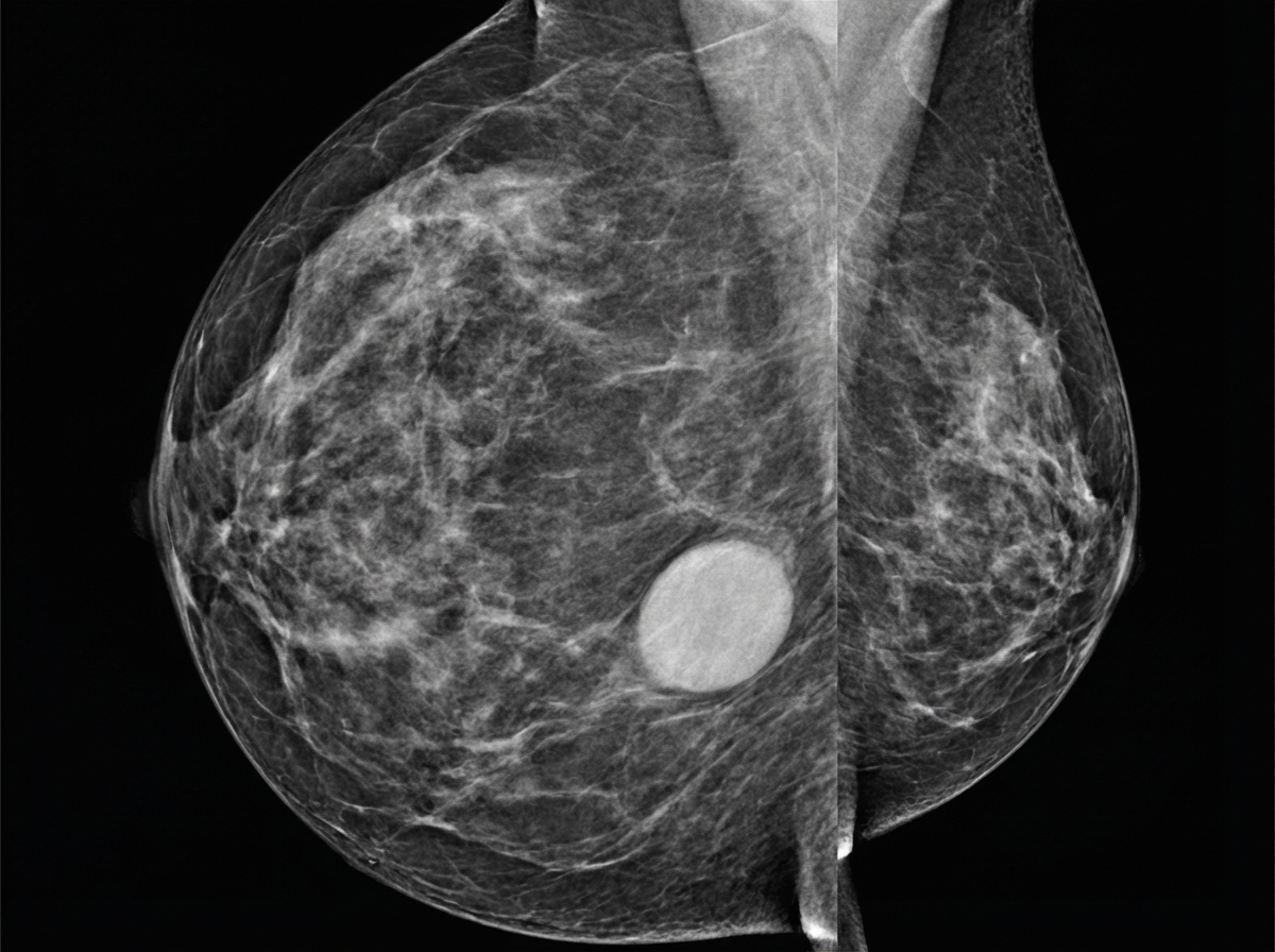
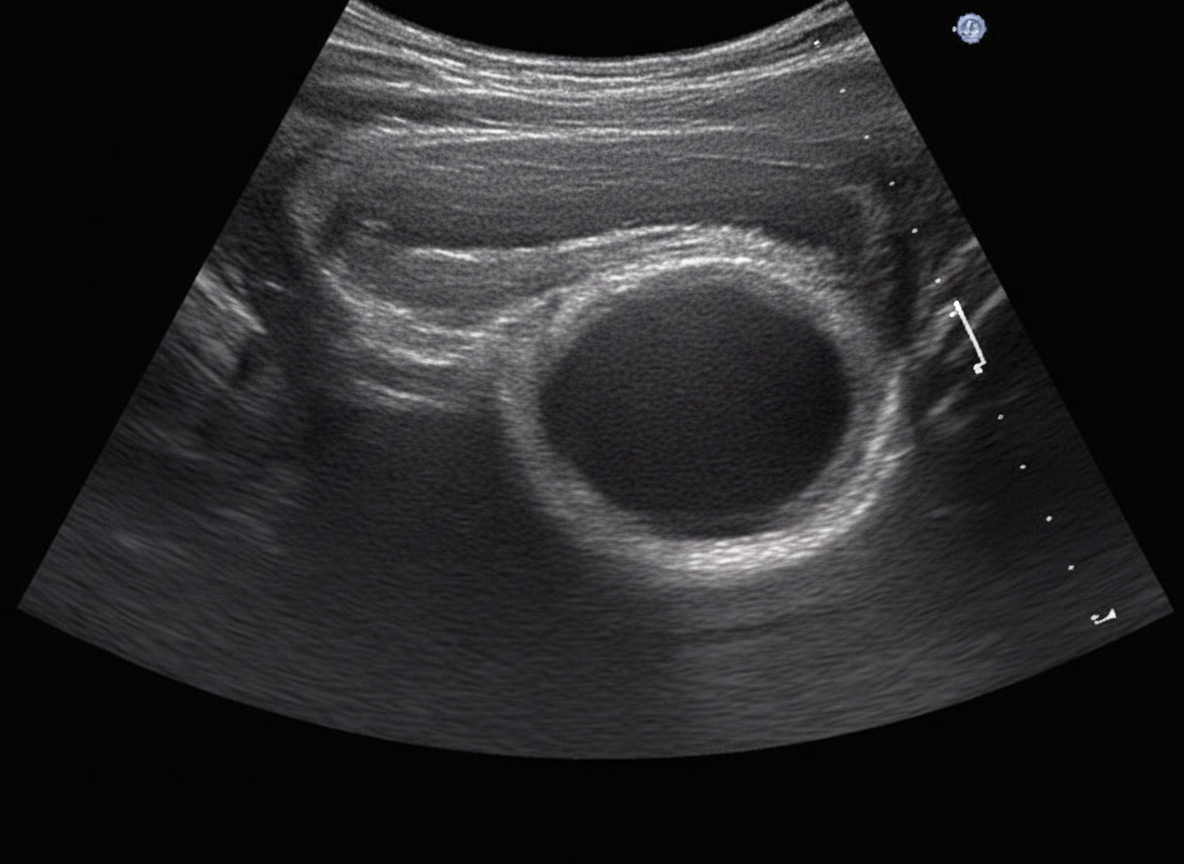
A 24-year-old female patient presents with bilateral breast mass. On physical examination this breast mass is hyper mobile and soft. The mammogram is shown below. What is the most possible diagnosis?

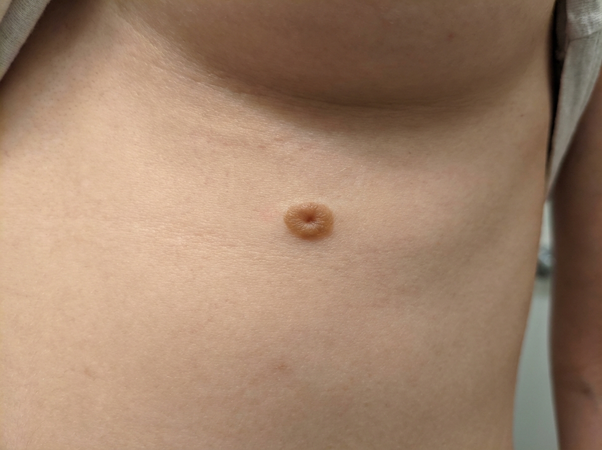
What is the diagnosis of the patient shown in the image? (Recent NEET Pattern 2016-17)

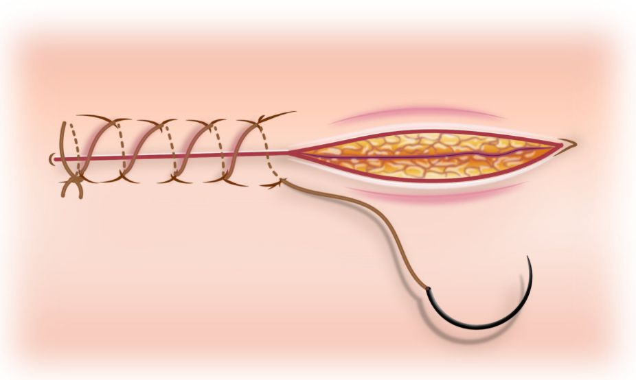
The surgeon attending has completed a modified radical mastectomy for a carcinoma breast patient. You have to suture the wound using subcuticular sutures. Which of the following sutures would you choose?

Which of the following is not an indication for surgery in the condition shown below?

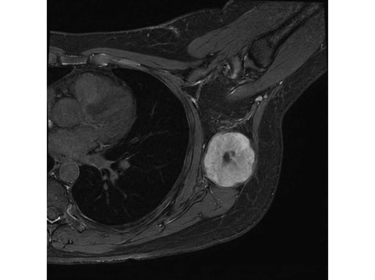
In the MRI breast shown below, 4 cm mass is present with no nodal metastasis. Which is the stage of breast cancer?

Explanation: ***Fibroadenoma*** - The patient's age (24 years old), bilateral location, **hypermobile** and **soft** mass on physical examination, together with the typical appearance on mammogram (well-circumscribed, oval lesion with calcifications) are characteristic of a **fibroadenoma**. - Fibroadenomas are **benign solid tumors** most common in young women, often appearing as distinct, movable lumps. *Breast cyst* - While breast cysts can be soft and mobile, they are typically **fluid-filled** and would appear as a **smooth, anechoic (dark) lesion on ultrasound**, not necessarily with dense calcifications seen on mammography. - Cysts are often associated with hormonal changes and can fluctuate in size; the described mass is solid on imaging evidence provided. *Ductal carcinoma in situ* - **Ductal carcinoma in situ (DCIS)** is a non-invasive form of breast cancer often presenting as **microcalcifications** in a linear or branching pattern, or as a mass with irregular margins. - The described mass is hypermobile and soft, and the mammographic appearance of the lesion in the image provided are not typical of DCIS which usually has spiculated margins and irregular shape. *Lobular carcinoma* - **Lobular carcinoma** (in situ or invasive) often presents as architectural distortion, an area of thickening, or as an insidious mass that is **poorly defined** and **difficult to detect** clinically or mammographically. - It is known for its "stealth" nature, often not forming a palpable lump or a distinct mass with the well-defined borders and hypermobility described. *Phyllodes tumor* - **Phyllodes tumors** are rare fibroepithelial tumors that can present as mobile breast masses in young women, but they typically grow **rapidly** and are often **larger** (>3-5 cm) at presentation. - The **bilateral** presentation and **typical mammographic appearance** favor fibroadenoma, as phyllodes tumors are usually **unilateral** and may show heterogeneous density with areas of necrosis or cystic change.
Explanation: ***Accessory nipple*** - The image distinctly shows small, pigmented lesions with a central indentation, consistently located along the **embryonic milk line**. This morphology and location are classic for **supernumerary nipples** or accessory nipples. - While they can vary in appearance, ranging from a simple pigmented macule to a fully developed nipple-areola complex, these lesions clearly fit the description of an accessory nipple. *Polymastia* - **Polymastia** refers to the presence of accessory breast tissue (glandular tissue) along the milk line, which may or may not include a nipple. - The key difference is that polymastia involves actual breast parenchyma, whereas the image shows only nipple structures without underlying breast tissue mass. - Both are congenital anomalies along the milk line, but the clinical presentation differs. *Hypertrophic nevus* - A **hypertrophic nevus** (or mole) is typically a raised, pigmented lesion, but it generally lacks the characteristic central depression or resemblance to nipple tissue seen in the image. - Nevi are common, but their appearance is usually uniform or nodular, not mimicking a breast structure. *Amazia* - **Amazia** refers to the congenital absence of breast tissue, while the nipple is present. - The image shows additional **nipple-like structures**, not the absence of breast tissue or nipples. *Melanoma* - **Melanoma** is a serious form of skin cancer characterized by irregular borders, asymmetry, varied color, and a changing diameter (**ABCDEs of melanoma**). - The lesions in the image appear symmetrical, well-defined, and uniform in color and shape, which are inconsistent with the highly irregular features of melanoma.
Explanation: ***A (Monocryl)*** - **Monocryl**, a **monofilament absorbable suture**, is ideal for subcuticular closure due to its smooth passage through tissue and minimal tissue reactivity. - Its absorbable nature ensures no removal is needed, which is cosmetically favorable for visible scars such as after a mastectomy. *B (Vicryl Plus)* - **Vicryl Plus** is an **absorbable braided suture**, which can have increased tissue drag and a higher risk of bacterial colonization compared to monofilament sutures, making it less ideal for subcuticular closure where a smooth passage and minimal foreign body reaction are preferred. - While absorbable, its braided structure might lead to more pronounced tissue reaction or track formation, negatively impacting the cosmetic outcome. *C (Mersilk)* - **Mersilk** is a **non-absorbable, natural braided silk suture**. It is not suitable for subcuticular closure because it would require removal, can cause significant tissue reaction, and is associated with a higher risk of infection and suture extrusion if left in place. - Its non-absorbable nature and braided multifilament structure make it inappropriate for buried layers where an absorbable, monofilament suture is generally preferred for optimal wound healing and cosmetic results. *D (Prolene)* - **Prolene** is a **non-absorbable monofilament polypropylene suture**. While it has smooth passage through tissue with minimal drag, it requires removal after healing, making it less desirable for subcuticular closure where an absorbable suture eliminates the need for suture removal. - Its non-absorbable nature means it would need to be removed 7-10 days post-operatively, adding patient discomfort and an additional clinic visit. *E (PDS)* - **PDS (polydioxanone)** is an **absorbable monofilament suture** with excellent tensile strength and a longer absorption profile (180-210 days) compared to Monocryl (90-120 days). - While suitable for subcuticular closure, its longer absorption time may be unnecessary for superficial skin closure, and Monocryl's faster absorption with adequate strength retention makes it more optimal for this specific application.
Explanation: ***Patient's decision*** - While patient preferences are crucial in medical decision-making, simply the **patient's decision alone is not a primary medical indication for surgery** in the absence of other objective criteria for a fibroadenoma. - Surgery for **fibroadenoma** is typically guided by clinical and radiological findings, not solely by patient request. *Size more than 5 cm* - A **fibroadenoma** with a size of **more than 5 cm** is generally considered a strong indication for surgical excision. - Large fibroadenomas can cause **cosmetic distortion**, discomfort, and may be harder to distinguish from malignant lesions, especially if they show rapid growth. *Complex type* - **Complex fibroadenomas** have features such as **cysts larger than 3 mm**, sclerosing adenosis, epithelial calcifications, or papillary apocrine metaplasia. - These features are associated with a slightly **increased risk of future breast cancer** and are often considered an indication for excision to rule out malignancy and for risk reduction. *Recurrence* - If a **fibroadenoma recurs** after previous excision, particularly if it grows rapidly or shows atypical features, surgical removal is indicated. - **Recurrence** suggests a persistent or potentially more aggressive benign process that warrants further investigation and management. *Rapid increase in size* - A **rapid increase in size** of a fibroadenoma is a clear indication for surgical excision. - Rapid growth raises concern for **phyllodes tumor** or other potentially aggressive lesions and warrants histopathological examination. - Serial measurements showing significant growth over a short period (typically doubling in size over 3-6 months) indicate the need for surgical intervention.
Explanation: ***Stage II*** - A tumor of **4 cm** with no nodal metastasis is classified as pT2, N0, M0. This falls under **Stage IIA** breast cancer according to the TNM staging system. - Stage II breast cancer typically includes tumors that are larger than 2 cm but not larger than 5 cm (T2N0), or smaller tumors with lymph node involvement. - The **4 cm size with N0 status** specifically defines Stage IIA. *Stage 0* - Stage 0 represents **ductal carcinoma in situ (DCIS)** or **lobular carcinoma in situ (LCIS)**, which are non-invasive breast cancers. - This case describes a **4 cm mass**, indicating an invasive tumor, not in-situ disease, ruling out Stage 0. *Stage I* - Stage I breast cancer includes tumors that are **2 cm or smaller** (T1) and have no lymph node involvement (N0). - Since the mass is **4 cm**, it exceeds the size criteria for Stage I. *Stage III* - Stage III breast cancer involves **larger tumors** (greater than 5 cm) or any tumor size with extensive lymph node involvement, or tumors invading the chest wall/skin. - While this mass is 4 cm, there is **no nodal metastasis**, which rules out Stage III. *Stage IV* - Stage IV breast cancer indicates the presence of **distant metastases** (M1), meaning the cancer has spread to other parts of the body. - The information provided clearly states **no nodal metastasis**, and no information about distant spread, therefore Stage IV is incorrect.
Axillary lymph node dissection
Practice Questions
Benign breast disease management
Practice Questions
Breast biopsy techniques
Practice Questions
Breast cancer staging and surgical management
Practice Questions
Breast conservation therapy
Practice Questions
Breast reconstruction options
Practice Questions
Hereditary breast cancer syndromes
Practice Questions
Inflammatory breast cancer
Practice Questions
Male breast cancer
Practice Questions
Mastectomy techniques and indications
Practice Questions
Nipple discharge evaluation
Practice Questions
Post-mastectomy complications
Practice Questions
Sentinel lymph node biopsy
Practice Questions
Get full access to all questions, explanations, and performance tracking.
Start For Free