
Abdominal emergencies — MCQs

On this page
A 62-year-old man presents to the office because of painless rectal bleeding for the past 3 months. He describes intermittent streaks of bright red blood on the toilet paper after wiping and blood on but not mixed within the stool. Occasionally, he has noted a small volume of blood within the toilet bowl, and he associates this with straining. For the past 2 weeks, he has noticed an 'uncomfortable lump' in his anus when defecating, which goes away by itself immediately afterwards. He says he has no abdominal pain, weight loss, or fevers. He is a well-appearing man that is slightly obese. Digital rectal examination shows bright red blood on the examination glove following the procedure. Anoscopy shows enlarged blood vessels above the pectinate line. Which of the following is the most likely cause?
A 26-year-old man presents to the emergency room with a complaint of lower abdominal pain that started about 5 hours ago. The pain was initially located around the umbilicus but later shifted to the right lower abdomen. It is a continuous dull, aching pain that does not radiate. He rates the severity of his pain as 7/10. He denies any previous history of similar symptoms. The vital signs include heart rate 100/min, respiratory rate 20/min, temperature 38.0°C (100.4°F), and blood pressure 114/77 mm Hg. On physical examination, there is severe right lower quadrant tenderness on palpation. Deep palpation of the left lower quadrant produces pain in the right lower quadrant. Rebound tenderness is present. The decision is made to place the patient on antibiotics and defer surgery. Two days later, his abdominal pain has worsened. Urgent computed tomography (CT) scan reveals new hepatic abscesses. The complete blood count result is given below: Hemoglobin 16.2 mg/dL Hematocrit 48% Leukocyte count 15,000/mm³ Neutrophils 69% Bands 3% Eosinophils 1% Basophils 0% Lymphocytes 24% Monocytes 3% Platelet count 380,000/mm³ Which of the following complications has this patient most likely experienced?
A 68-year-old man presents with a 3-month history of difficulty starting urination, weak stream, and terminal dribbling. The patient has no history of serious illnesses and is not under any medications currently. The patient’s father had prostate cancer at the age of 58 years. Vital signs are within normal range. Upon examination, the urinary bladder is not palpable. Further examination reveals normal anal sphincter tone and a bulbocavernosus muscle reflex. Digital rectal exam (DRE) shows a prostate size equivalent to 2 finger pads with a hard nodule and without fluctuance or tenderness. The prostate-specific antigen (PSA) level is 5 ng/mL. Image-guided biopsy indicates prostate cancer. MRI shows tumor confined within the prostate. Radionuclide bone scan reveals no abnormalities. Which of the following interventions is the most appropriate next step in the management of this patient?
A 43-year-old woman is brought to the emergency department for evaluation of worsening abdominal pain that suddenly started 2 hours ago. The patient also has nausea and has vomited twice. She has hypothyroidism, systemic lupus erythematosus, major depressive disorder, and chronic right knee pain. Current medications include levothyroxine, prednisone, fluoxetine, naproxen, and a chondroitin sulfate supplement. She appears distressed. Her temperature is 37.9°C (100.2°F), pulse is 101/min, and blood pressure is 115/70 mm Hg. Examination shows a rigid abdomen with rebound tenderness; bowel sounds are hypoactive. Laboratory studies show a leukocyte count of 13,300/mm3 and an erythrocyte sedimentation rate of 70 mm/h. An x-ray of the chest is shown. Which of the following is the most appropriate next step in management?
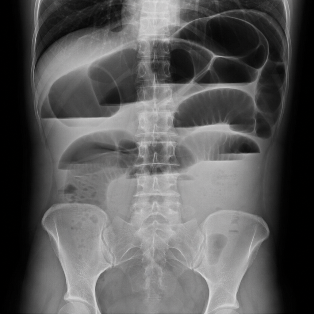
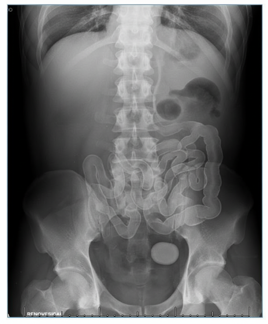
An 82-year-old woman visits her primary care provider complaining of a vague cramping pain on the right side of her abdomen for the past 6 hours. She is also nauseated and had an episode of vomiting earlier today and two episodes yesterday. Past medical history includes third-degree heart block, gastroesophageal reflux disease, hypertension, hypothyroidism and chronic cholecystitis with cholelithiasis. She is not a good candidate for cholecystectomy due to cardiac disease and is treated with analgesics and ursodeoxycholic acid. Her medications include chlorthalidone, omeprazole, levothyroxine, and occasional naproxen for pain. Vitals are normal. A supine abdominal X-ray reveals air in the gallbladder and biliary tree, small bowel obstruction, and a large radiolucent gallstone impacted in the small bowel. What is the most likely diagnosis?
A 29-year-old man presents to the emergency room with severe abdominal pain. He states that for the entire day, he has had pain in his lower right abdomen in addition to a loss of appetite accompanied by nausea and vomiting. His temperature is 101.3°F (38.5°C), blood pressure is 125/98 mmHg, pulse is 78/min, and respirations are 15/min. On physical examination, he exhibits increased abdominal pain in his right lower quadrant upon deep palpation of the left lower quadrant. What is the next step in the management of this patient?
A 72-year-old man is brought to the emergency department with increasing fever and abdominal pain over the past week. The pain is constant and limited to the lower right part of his abdomen. He has nausea but no vomiting or diarrhea. His past medical history is unremarkable for any serious illnesses. He takes acetaminophen for knee arthritis. He is fully alert and oriented. His temperature is 39.5°C (103.1°F), pulse is 89/min, respirations are 15/min, and blood pressure is 135/70 mm Hg. Abdominal examination shows a tender mass in the right lower quadrant. CT shows obstruction of the appendiceal neck with a fecalith and the appendiceal tip leading to an irregular walled-off fluid collection. Stranding of the surrounding fat planes is also noted. Intravenous hydration is initiated. Which of the following is the most appropriate next step in management?
A 66-year-old man comes to the physician because of yellowish discoloration of his eyes and skin, abdominal discomfort, and generalized fatigue for the past 2 weeks. He has had dark urine and pale stools during this period. He has had a 10-kg (22-lb) weight loss since his last visit 6 months ago. He has hypertension. He has smoked one pack of cigarettes daily for 34 years. He drinks three to four beers over the weekends. His only medication is amlodipine. His temperature is 37.3°C (99.1°F), pulse is 89/min, respirations are 14/min, and blood pressure is 114/74 mm Hg. Examination shows jaundice of the sclera and skin and excoriation marks on his trunk and extremities. The lungs are clear to auscultation. The abdomen is soft and nontender. The remainder of the examination shows no abnormalities. Laboratory studies show: Hemoglobin 12 g/dL Leukocyte count 5,000/mm3 Platelet count 400,000/mm3 Serum Urea nitrogen 28 mg/dL Creatinine 1.2 mg/dL Bilirubin Total 7.0 mg/dL Direct 5.5 mg/dL Alkaline phosphatase 615 U/L Aspartate aminotransferase (AST, GOT) 170 U/L Alanine aminotransferase (ALT, GPT) 310 U/L γ-Glutamyltransferase (GGT) 592 U/L (N = 5–50 U/L) An ultrasound shows extrahepatic biliary dilation. A CT scan of the abdomen shows a 2.5-cm (1-in) mass in the head of the pancreas with no abdominal lymphadenopathy. The patient undergoes biliary stenting. Which of the following is the most appropriate next step in the management of this patient?
A 59-year-old woman presents to the family medicine clinic with a lump in her breast for the past 6 months. She states that she has been doing breast self-examinations once a month. She has a medical history significant for generalized anxiety disorder and systemic lupus erythematosus. She takes sertraline and hydroxychloroquine for her medical conditions. The heart rate is 102/min, and the rest of the vital signs are stable. On physical examination, the patient appears anxious and tired. Her lungs are clear to auscultation bilaterally. Capillary refill is 2 seconds. There is no axillary lymphadenopathy present. Palpation of the left breast reveals a 2 x 2 cm mass. What is the most appropriate next step given the history of the patient?
A 68-year-old man presents for a screening ultrasound scan. He has been feeling well and is in his usual state of good health. His medical history is notable for mild hypertension and a 100-pack-year tobacco history. He has a blood pressure of 128/86 and heart rate of 62/min. Physical examination is clear lung sounds and regular heart sounds. On ultrasound, an infrarenal aortic aneurysm of 4 cm in diameter is identified. Which of the following is the best initial step for this patient?
Practice by Chapter
Acute appendicitis
Practice Questions
Acute cholecystitis
Practice Questions
Small bowel obstruction
Practice Questions
Large bowel obstruction
Practice Questions
Mesenteric ischemia
Practice Questions
Gastrointestinal perforation
Practice Questions
Abdominal compartment syndrome
Practice Questions
Acute pancreatitis management
Practice Questions
Diverticulitis
Practice Questions
Incarcerated/strangulated hernias
Practice Questions
Non-operative management principles
Practice Questions
Diagnostic approach to acute abdomen
Practice Questions
Damage control surgery
Practice Questions
Want unlimited practice?
Get full access to all questions, explanations, and performance tracking.
Scan to download app