Among the following options, what is the most common known single-factor cause of congenital anomalies?
Chloride level in sweat is used in the diagnosis of which disease?
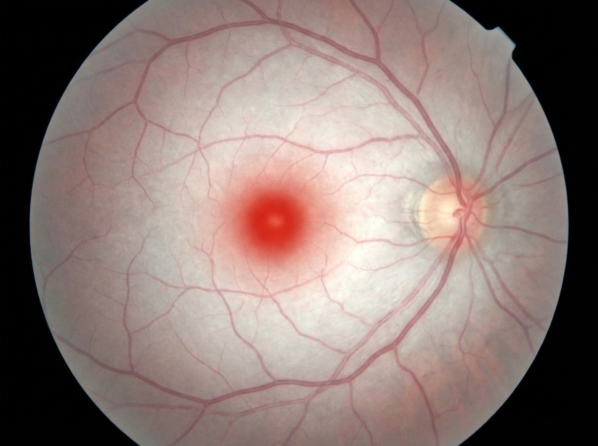
A 3-year-old boy presents with developmental delays and an inability to walk. The fundoscopy image is given below. What is the most likely diagnosis?

Which of the following statements about Fragile X syndrome is true?
Risk of genetic diseases in consanguineous marriage between first cousins?
What is the current medical terminology used to refer to Down syndrome?
Which of the following statements is MOST accurate regarding ataxia telangiectasia?
Which of the following is a characteristic feature of DiGeorge syndrome?
Alagille syndrome - which of the following statements is false?
Which of the following is NOT a feature of CHARGE syndrome?
Explanation: ***Chromosomal aberrations*** - Chromosomal abnormalities, such as **aneuploidies** and **structural rearrangements**, are the most frequent single-factor cause of **congenital anomalies**, accounting for a significant proportion of birth defects. - Examples include **Down syndrome** (trisomy 21), **Turner syndrome** (XO), and **Klinefelter syndrome** (XXY), which lead to distinct clinical presentations and developmental issues. *Maternal infections* - While maternal infections like **TORCH infections (Toxoplasmosis, Other [syphilis, varicella-zoster, parvovirus B19], Rubella, Cytomegalovirus, and Herpes simplex virus)** can cause congenital anomalies, they represent a smaller proportion of overall birth defects compared to chromosomal aberrations. - The type of anomaly depends on the specific pathogen and the timing of infection during gestation. *Drugs* - **Teratogenic drugs**, such as **thalidomide** or **valproic acid**, can cause severe congenital anomalies, but they are responsible for a relatively small percentage of all birth defects. - The impact depends on the drug, dose, and the stage of fetal development during exposure. *Irradiation* - **Ionizing radiation exposure** during pregnancy, especially during critical periods of organogenesis, can lead to congenital anomalies, developmental delays, and other adverse outcomes. - However, significant teratogenic exposure to radiation is **uncommon** in most populations, making it a less frequent cause compared to other factors. *Multifactorial inheritance* - **Multifactorial disorders** result from the interaction of multiple genes and environmental factors, accounting for the **majority** of congenital anomalies overall (e.g., neural tube defects, cleft lip/palate, congenital heart defects). - However, the question asks for **single-factor** causes, and multifactorial conditions by definition involve multiple contributing factors, not a single identifiable cause.
Explanation: ***Cystic fibrosis*** - **Cystic fibrosis** is characterized by a defect in the **CFTR protein**, leading to impaired chloride transport in epithelial cells. - This defect results in abnormally high salt content in sweat, making the **sweat chloride test** the gold standard for diagnosis. *Phenylketonuria* - **Phenylketonuria (PKU)** is a metabolic disorder involving the inability to metabolize **phenylalanine**, leading to its accumulation in the blood. - Diagnosis is typically made via **newborn screening** using blood tests, not sweat chloride levels. *Gaucher's disease* - **Gaucher's disease** is a lysosomal storage disorder caused by a deficiency in the enzyme **beta-glucosidase**, leading to lipid accumulation. - Diagnosis involves enzyme assays from blood or tissue samples, or genetic testing, not sweat chloride analysis. *Osteogenesis imperfecta* - **Osteogenesis imperfecta** is a genetic disorder of **collagen synthesis**, primarily characterized by bone fragility, blue sclerae, and hearing loss. - Diagnosis is based on clinical features, imaging, and genetic testing, with no relevance to sweat chloride levels. *Tay-Sachs disease* - **Tay-Sachs disease** is a lysosomal storage disorder caused by deficiency of **hexosaminidase A**, leading to accumulation of GM2 ganglioside in neurons. - Diagnosis is made through **enzyme assay** or **genetic testing**, not sweat chloride measurement.
Explanation: ***Tay-Sachs disease*** - The image shows a **cherry-red spot** in the macula, a classic ocular finding in Tay-Sachs disease caused by **ganglioside accumulation** in retinal ganglion cells. - The clinical presentation of **developmental delays** and inability to walk in a 3-year-old boy is consistent with the neurodegenerative course of Tay-Sachs disease. - Tay-Sachs is **more common** than Niemann-Pick disease and classically presents with this triad: cherry-red spot, developmental regression, and hypotonia. *Niemann-Pick disease* - Niemann-Pick disease (especially Type A) can also present with a **cherry-red spot** and developmental delay, making it a close differential. - However, Niemann-Pick typically presents earlier (infancy) with more prominent **hepatosplenomegaly** and **sphingomyelin accumulation**, which are not mentioned in this case. - The age of presentation (3 years) and classic fundoscopic finding favor Tay-Sachs over Niemann-Pick. *Hunter syndrome* - Hunter syndrome is a lysosomal storage disorder characterized by **coarse facial features**, hepatosplenomegaly, and skeletal abnormalities, but typically **does not present with a cherry-red spot**. - While developmental delay can occur, the absence of a cherry-red spot and presence of other physical stigmata differentiate it from Tay-Sachs. *Hurler syndrome* - Hurler syndrome is another mucopolysaccharidosis with features similar to Hunter syndrome, including **skeletal deformities**, coarse facial features, and **corneal clouding**, but **not a cherry-red spot**. - The significant developmental regression and the specific fundoscopic finding point away from Hurler syndrome. *Gaucher disease* - Gaucher disease is characterized by **hepatosplenomegaly**, bone pain, and neurologic involvement in some forms, but it is **not typically associated with a cherry-red spot** in the fundus. - While developmental delay can be present, the absence of the pathognomonic ocular finding makes Tay-Sachs a more likely diagnosis.
Explanation: ***Males have IQ 20-40*** - Males with Fragile X syndrome typically exhibit **moderate to severe intellectual disability**, with IQ scores commonly ranging from 20 to 40. - This intellectual impairment is a hallmark feature due to the **loss of FMRP protein function** in brain development. *Triple nucleotide CAG Sequence mutation* - Fragile X syndrome is caused by an expansion of a **CGG triplet repeat** (not CAG) in the **FMR1 gene**. - This CGG repeat expansion leads to **hypermethylation** and silencing of the FMR1 gene. *25-30% Female carriers have intellectual disability* - While some female carriers may experience milder symptoms or learning disabilities, only about **50% of female carriers have intellectual disability**, typically mild, not 25-30%. - The variable expressivity in females is due to **X-inactivation**, where the unaffected X chromosome can partially compensate. *Gain of function mutation* - Fragile X syndrome is caused by a **loss of function mutation** in the **FMR1 gene** due to the CGG repeat expansion. - This leads to the absence or severe reduction of the **fragile X mental retardation protein (FMRP)**, which is crucial for synaptic development and function. *Autosomal dominant inheritance pattern* - Fragile X syndrome follows an **X-linked dominant inheritance pattern** (not autosomal dominant). - Males are more severely affected than females because they have only one X chromosome, while females have two X chromosomes (one may be unaffected).
Explanation: ***4-8%*** - The risk of genetic diseases in offspring from a **first-cousin consanguineous marriage** is generally estimated to be **two- to threefold higher than the baseline risk** in the general population. - While the general population risk for a serious birth defect or genetic disease is around 3-4%, this increased risk typically places the range for first cousins at **4-8%**. *1-2%* - This percentage represents a **lower risk than what is typically observed** with first-cousin consanguinity. - The baseline risk for genetic disease in the general population is usually around 3-4%, and consanguineous unions significantly increase this risk. *2-4%* - This represents approximately the **baseline population risk** without consanguinity, not the increased risk seen with first-cousin marriages. - Students may incorrectly assume that first-cousin marriage does not significantly elevate the baseline genetic disease risk. *8-10%* - While there is an increased risk, this range is generally considered **higher than the average estimated risk** for first-cousin consanguineous marriages, which is typically quoted at 4-8%. - This higher range might be seen in cases of **more distant consanguinity** or in populations with a **higher prevalence of specific recessive conditions**. *12-14%* - This magnitude of risk is usually associated with **closer degrees of consanguinity**, such as uncle-niece or half-sibling relationships, rather than first cousins. - The **coefficient of inbreeding** for first cousins is 1/16, which carries a lower risk than these closer relationships.
Explanation: ***Trisomy 21*** - This is the **correct and most specific medical terminology** for Down syndrome, indicating the presence of an extra copy of chromosome 21. - It directly describes the **genomic abnormality** that causes the syndrome. *Trisomy 18* - This refers to **Edwards syndrome**, a different chromosomal disorder caused by an extra copy of chromosome 18. - While also a trisomy, it is clinically distinct from Down syndrome with different characteristic features. *Trisomy 13* - This refers to **Patau syndrome**, another chromosomal disorder caused by an extra copy of chromosome 13. - Like Trisomy 18, it is a distinct trisomy condition but not the correct terminology for Down syndrome. *Chromosomal disorder* - This is a **broad category** that includes Down syndrome but is not the specific medical term. - Many conditions, like Turner syndrome or Klinefelter syndrome, are also chromosomal disorders, but they are genetically and clinically distinct. *Genetic disorder* - This is an **even broader category** encompassing any disease caused by a genetic abnormality. - While Down syndrome is indeed a genetic disorder, this term is not specific enough to identify it.
Explanation: ***Increase in AFP*** - Ataxia telangiectasia is associated with elevated **alpha-fetoprotein (AFP)** levels due to impaired liver function and cellular responses. - This condition is linked with immunodeficiency, leading to abnormal AFP metabolism in affected individuals. *Autosomal dominant* - Ataxia telangiectasia follows an **autosomal recessive** inheritance pattern, not dominant [1]. - It is caused by mutations in the **ATM gene**, which is involved in DNA repair. *Increases the risk of squamous cell carcinoma* - While ataxia telangiectasia patients have a higher risk of **lymphoma** and **leukemia**, they do not have a significant increased risk for **squamous cell carcinoma**. - The malignancy risk is more associated with **lymphoid tissues**, rather than skin cancers. *None of above* - This option is incorrect as the **increased AFP** in ataxia telangiectasia is a well-documented clinical feature. - Hence, stating that "none of the above" is true is inaccurate. **References:** [1] Kumar V, Abbas AK, et al.. Robbins and Cotran Pathologic Basis of Disease. 9th ed. The Central Nervous System, pp. 1300-1301.
Explanation: ***Hypoparathyroidism leading to tetany*** - DiGeorge syndrome is characterized by **thymic and parathyroid hypoplasia or aplasia**, due to a deletion on **chromosome 22q11.2**. - **Hypoparathyroidism** results in **hypocalcemia**, which can manifest as **tetany** (muscle spasms due to low calcium). - This is a classic and highly characteristic feature of DiGeorge syndrome. *Eczema* - While patients with immune deficiencies, including some with DiGeorge syndrome, can be prone to skin infections, **eczema** is not a primary or characteristic feature used for diagnosis. - Eczema is more commonly associated with other immune disorders like **Wiskott-Aldrich syndrome** or severe combined immunodeficiency, but not typically DiGeorge syndrome. *Normal B-cell function with T-cell deficiency* - DiGeorge syndrome is characterized by **T-cell deficiency** due to thymic abnormalities; however, B-cell function can be affected indirectly or through other mechanisms, making "normal B-cell function" not universally accurate. - While **T-cell deficiency** is a hallmark, the precise nature of B-cell involvement can vary, and often B-cell function is also impaired, albeit less directly. *Partial T-cell deficiency* - Although it's often described as a **partial T-cell deficiency**, a more precise characteristic is the **thymic hypoplasia or aplasia**, which directly impairs T-cell development. - The degree of T-cell deficiency can vary from mild to severe, but identifying the underlying cause of **hypoparathyroidism** is a more specific diagnostic feature. *Hyperparathyroidism with hypercalcemia* - This is the **opposite** of what occurs in DiGeorge syndrome. - DiGeorge syndrome presents with **hypoparathyroidism and hypocalcemia**, not hyperparathyroidism. - Hyperparathyroidism would cause elevated calcium levels, which is not seen in this condition.
Explanation: ***Autosomal Recessive Disease*** - Alagille syndrome is an **autosomal dominant** disorder, meaning only one copy of the mutated gene is needed to cause the condition. - The mode of inheritance is critical for genetic counseling and understanding disease patterns within families. *Mutation in JAG 1 And Notch2 gene are seen* - Alagille syndrome is primarily caused by mutations in the **JAG1 gene** (94% of cases), which encodes a ligand for the Notch receptor. - While less common, mutations in the **NOTCH2 gene** can also lead to Alagille syndrome, as both genes are part of the Notch signaling pathway essential for development. *Does not cause Autoimmune hepatitis* - Alagille syndrome is a multisystem disorder characterized by **cholestasis** due to bile duct paucity, which is distinct from autoimmune hepatitis. - Although both involve liver pathology, autoimmune hepatitis is an inflammatory process mediated by the immune system, and it is not a direct feature or cause of Alagille syndrome. *Valvular anomalies of heart are seen* - **Pulmonary artery stenosis** (peripheral or main) is the most common cardiac anomaly found in Alagille syndrome, occurring in over 90% of affected individuals. - Other cardiac defects, such as **tetralogy of Fallot**, are also observed, highlighting the syndrome's impact on cardiovascular development. *Posterior embryotoxon is a characteristic ocular finding* - **Posterior embryotoxon** (a prominent, anteriorly displaced Schwalbe line) is present in approximately **90% of patients** with Alagille syndrome. - This ocular finding is one of the five major diagnostic criteria for the syndrome, along with cholestasis, cardiac defects, skeletal abnormalities, and characteristic facial features.
Explanation: ***Esophageal Atresia*** - While other **tracheoesophageal abnormalities** can occur in CHARGE syndrome, **esophageal atresia** is not typically considered one of the core diagnostic criteria. - The acronym CHARGE stands for a specific set of features, and esophageal atresia is not directly included, unlike **choanal atresia**. *Eye Coloboma* - **Coloboma** of the eye, particularly the iris or retina, is a **major diagnostic feature** of CHARGE syndrome. - This congenital malformation results from incomplete closure of the **choroid fissure** during development. *Congenital heart disease* - Various types of **congenital heart defects**, such as **tetralogy of Fallot** or an **atresia of the aortic valve**, are common in CHARGE syndrome. - These cardiac anomalies are a **major diagnostic criterion** and significantly impact patient management. *Choanal Atresia* - **Choanal atresia** represents the "A" in the CHARGE acronym and is a **major diagnostic criterion**. - This condition involves blockage of the nasal passages and is one of the most characteristic features of the syndrome. *Urinary tract defects* - **Genital and urinary abnormalities** are frequently observed in individuals with CHARGE syndrome. - These can include **hypoplastic genitalia** and **kidney abnormalities**, classifying as a **minor diagnostic feature**.
Chromosomal Disorders
Practice Questions
Disorders of Amino Acid Metabolism
Practice Questions
Disorders of Carbohydrate Metabolism
Practice Questions
Disorders of Lipid Metabolism
Practice Questions
Genetic Counseling
Practice Questions
Genetic Testing in Pediatrics
Practice Questions
Inborn Errors of Metabolism
Practice Questions
Lysosomal Storage Diseases
Practice Questions
Mitochondrial Disorders
Practice Questions
Multifactorial Inheritance Disorders
Practice Questions
Newborn Screening
Practice Questions
Single Gene Disorders
Practice Questions
Get full access to all questions, explanations, and performance tracking.
Start For Free