Phenotypic expression of a gene depending on the parent of origin is referred to as:
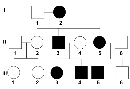
All of the following are tests done for Turner mosaic screening except?
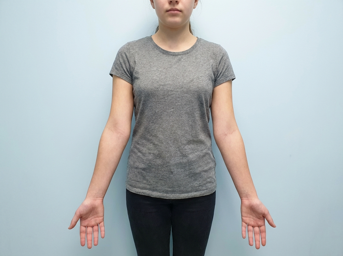
A teenage girl presents with a history of amenorrhea. Local examination is shown in the image. What karyotype analysis would you consider for further evaluation?

A baby's blood group was determined as O negative. Which of the following blood groups can the baby's mother or father not have?
Which of the following techniques is used for the detection of variations in DNA sequence and gene expression?
34 week primigravida punjabi khatri comes with history of consanguineous marriage, with history of repeated blood transfusion to her sibling since 8 months of age. The first diagnostic test is -
A 4-year-old boy is brought to a pediatrician by his parents for a consultation after his teacher complained about his inability to focus or make friends at school. They mention that the boy does not interact well with others at home, school, or daycare. On physical examination, his vital signs are stable with normal weight, height, and head circumference for his age and sex. His general examination and neurologic examination are completely normal. A recent audiological evaluation shows normal hearing, and intellectual disability has been ruled out by a clinical psychologist. Which of the following investigations is indicated as part of his diagnostic evaluation at present?
What is the chance of a child having cystic fibrosis if both parents are carriers of the disease?
A 34-year-old gravida 2 para 1 woman at 16 weeks gestation presents for prenatal care. Her prenatal course has been uncomplicated. She takes no medications besides her prenatal vitamin which she takes every day, and she has been compliant with routine prenatal care. She has a 7-year-old daughter who is healthy. The results of her recent quadruple screen are listed below: AFP: Low hCG: Low Estriol: Low Inhibin-A: Normal Which of the following is the most appropriate next step to confirm the diagnosis?
A 25-year-old man with a genetic disorder presents for genetic counseling because he is concerned about the risk that any children he has will have the same disease as himself. Specifically, since childhood he has had difficulty breathing requiring bronchodilators, inhaled corticosteroids, and chest physiotherapy. He has also had diarrhea and malabsorption requiring enzyme replacement therapy. If his wife comes from a population where 1 in 10,000 people are affected by this same disorder, which of the following best represents the likelihood a child would be affected as well?
Explanation: ***Genomic imprinting (parent-of-origin gene expression)*** - **Genomic imprinting** is an epigenetic phenomenon where gene expression is dependent on whether the gene was inherited from the mother or the father. - This results in monoallelic expression of specific genes, with only one copy (maternal or paternal) being active. *Mosaic genetic variation* - **Mosaicism** refers to the presence of two or more populations of genetically different cells in one individual, all derived from a single zygote. - This typically arises from a somatic mutation during development, not from differential expression based on parental origin. *Nonpenetrance of genotype* - **Nonpenetrance** occurs when individuals carrying a disease-causing genotype do not express the associated phenotype. - This concept relates to the presence or absence of a phenotype, not the differential expression based on parental origin. *Genetic anticipation* - **Genetic anticipation** is the phenomenon where the symptoms of a genetic disorder become more severe and/or appear at an earlier age in successive generations. - This is commonly observed in disorders caused by expansions of trinucleotide repeats, such as Huntington's disease, and is distinct from parent-of-origin gene expression.
Explanation: ***Serum FSH*** - **Serum Follicle-Stimulating Hormone (FSH)** levels are used to assess ovarian function and can be elevated in conditions like Turner syndrome due to **gonadal dysgenesis**, but it is a **functional test**, not a screening tool for mosaicism. - While elevated FSH is a clinical feature of Turner syndrome, it does not directly screen for the chromosomal mosaicism itself. *Karyotype* - **Karyotyping** is the **gold standard** for diagnosing Turner syndrome and its mosaics by visualizing the entire set of chromosomes [1]. - It can identify various forms of mosaicism involving the X chromosome, where some cells have 45,XO and others have 46,XX or other variations [1]. *FISH* - **Fluorescence in situ hybridization (FISH)** is a molecular cytogenetic technique used to detect specific chromosomal abnormalities, including those associated with Turner mosaicism. - It uses DNA probes that bind to specific regions of the X chromosome, allowing for the rapid detection of **aneuploidy** or deletions that might indicate mosaicism [2]. *Buccal smear* - A **buccal smear**, historically used for **Barr body** analysis, can provide an initial screening for X chromosome abnormalities. - The presence of Barr bodies (inactive X chromosomes) can help differentiate between 45,XO (no Barr body) and mosaic variants like 45,XO/46,XX (variable number of Barr bodies). **References:** [1] Cross SS. Underwood's Pathology: A Clinical Approach. 6th ed. (Basic Pathology) introduces the student to key general principles of pathology, both as a medical science and as a clinical activity with a vital role in patient care. Part 2 (Disease Mechanisms) provides fundamental knowledge about the cellular and molecular processes involved in diseases, providing the rationale for their treatment. Part 3 (Systematic Pathology) deals in detail with specific diseases, with emphasis on the clinically important aspects., pp. 54-55. [2] Kumar V, Abbas AK, et al.. Robbins and Cotran Pathologic Basis of Disease. 9th ed. Genetic Disorders, pp. 186-187.
Explanation: ***45 XO*** - The image shows a **webbed neck** and **short stature** (suggested by the overall body proportions typically associated with Ullrich-Turner Syndrome), alongside primary amenorrhea, which are classic features of **Turner Syndrome**. - **Turner Syndrome** is a chromosomal disorder characterized by the absence of all or part of one X chromosome in females, resulting in a **45, XO karyotype**. *46 XY* - This karyotype indicates a **phenotypic male** with normal male chromosomal constitution. - Individuals with this karyotype would not typically present with **primary amenorrhea** as they do not have a uterus. *46 XX* - This is the **normal female karyotype**, and while a female with this karyotype could experience amenorrhea (e.g., due to Asherman's syndrome or PCOS), the physical features associated with the image (like webbed neck) are not consistent. - This option does not explain the **physical stigmata** often seen in genetic causes of primary amenorrhea, such as in Turner syndrome. *47 XXY* - This karyotype is characteristic of **Klinefelter Syndrome**, which affects males and is associated with hypogonadism and gynecomastia. - It would not be found in a female patient presenting with **amenorrhea** and the physical features shown in the image. *47 XXX* - This karyotype represents **Triple X Syndrome** (Trisomy X), which affects females and typically presents with **normal female appearance** and often normal fertility. - While some individuals may have menstrual irregularities, the **distinctive physical features** shown in the image (webbed neck, short stature) are not characteristic of Triple X syndrome, which usually lacks specific dysmorphic features.
Explanation: ***AB Negative*** - A parent with **AB blood type** cannot have an O blood type child because an individual with AB blood type only has A and B alleles to pass on (i.e., *I<sup>A</sup>I<sup>B</sup>* genotype). - For a child to have **O blood type**, they must inherit the *i* allele from both parents (*ii* genotype), which is impossible if one parent is AB. *A Positive* - A parent with **A positive blood type** can have an O negative child if their genotype is *I<sup>A</sup>i* for A/B/O and *Rr* for Rh factor. - The child would inherit the *i* allele from this parent and the *r* allele for Rh, along with the same from the other parent. *B Positive* - Similar to A positive, a parent with **B positive blood type** can have an O negative child if their genotype is *I<sup>B</sup>i* and *Rr*. - The child would inherit the *i* allele from this parent and the *r* allele for Rh, along with the same from the other parent. *O positive* - A parent with **O positive blood type** can certainly have an O negative child; their genotype would be *ii* for A/B/O and *Rr* for Rh factor. - The child would inherit the *i* allele from this parent and the *r* allele for Rh.
Explanation: ***Microarray*** - **Microarrays** are designed to detect thousands of DNA or RNA sequences simultaneously, making them ideal for analyzing **gene expression profiles** and identifying **sequence variations** like SNPs. - They involve hybridizing labeled sample DNA/RNA to probes fixed on a solid surface, with the intensity of hybridization indicating the presence or abundance of specific sequences. *Northern blot* - The **Northern blot** technique is primarily used to study **gene expression** by detecting specific **RNA sequences** in a sample. - It does not directly analyze DNA sequence variations. *Southern blot* - The **Southern blot** is a molecular biology method used to detect specific **DNA sequences** in DNA samples. - While it can identify large-scale DNA rearrangements or deletions, it is not optimized for simultaneous detection of multiple gene expression levels or subtle sequence variations. *Western blot* - The **Western blot** is used to detect specific **proteins** in a sample. - It analyzes protein expression levels and modifications and is not designed for the detection of DNA sequence variations or gene expression at the RNA level.
Explanation: ***Hb electrophoresis*** - The patient's history of **consanguineous marriage**, a sibling requiring **repeated blood transfusions** since 8 months of age, and Punjabi Khatri ethnicity strongly suggest a **hemoglobinopathy**, likely **beta-thalassemia major or intermedia**. - **Hemoglobin electrophoresis** is the traditional gold standard for definitive diagnosis of various hemoglobin variants and thalassemia types, identifying and characterizing abnormal hemoglobin patterns (e.g., elevated HbF, HbA2). - It remains a primary diagnostic test for hemoglobinopathies, particularly useful for pattern recognition of various thalassemia syndromes. *HPLC* - **High-performance liquid chromatography (HPLC)** is an equally valid and increasingly preferred method for diagnosing hemoglobinopathies, offering automated, precise quantification of hemoglobin fractions (HbA, HbA2, HbF). - In modern practice, HPLC is often used as a first-line screening tool due to its accuracy, reproducibility, and ability to provide quantitative data crucial for thalassemia diagnosis. - Both HPLC and Hb electrophoresis are acceptable diagnostic approaches; the choice between them depends on laboratory availability and practice patterns. For this 2013 exam, Hb electrophoresis was considered the traditional first diagnostic test. *Blood smear* - A **peripheral blood smear** would show morphological changes like **microcytic hypochromic red blood cells**, **target cells**, **anisopoikilocytosis**, and **nucleated RBCs**, which are suggestive of thalassemia. - These findings are indicative but non-specific and require confirmatory tests like hemoglobin electrophoresis or HPLC to identify the specific hemoglobin disorder and establish a definitive diagnosis. *Bone marrow* - A **bone marrow** examination would show **erythroid hyperplasia** due to increased ineffective erythropoiesis in thalassemia but is an invasive procedure and not the initial diagnostic test for hemoglobinopathies. - It provides details about cellularity and maturation but does not directly identify hemoglobin abnormalities, making it unsuitable as the first diagnostic step in suspected hemoglobinopathies.
Explanation: ***Autism spectrum disorder screening and developmental assessment*** - The clinical presentation (inability to focus, difficulty making friends, poor social interaction across multiple settings) is **highly suggestive of Autism Spectrum Disorder (ASD)**. - After ruling out **hearing impairment and intellectual disability**, the next appropriate step is **formal ASD screening using validated tools** such as the **Modified Checklist for Autism in Toddlers (M-CHAT)**, **Autism Diagnostic Observation Schedule (ADOS)**, or **Autism Diagnostic Interview-Revised (ADI-R)**. - According to **AAP guidelines**, when developmental concerns suggestive of ASD are identified, formal screening and comprehensive developmental assessment are **essential components of the diagnostic evaluation**. - ASD diagnosis is primarily **clinical**, based on standardized screening tools and developmental assessments, not neuroimaging or electrophysiological studies. *No further testing is needed* - This is **incorrect** because the patient has not yet undergone **formal ASD-specific screening and developmental assessment**. - While hearing and intellectual disability have been ruled out, **diagnostic confirmation of ASD** requires structured evaluation using validated assessment tools. - Simply observing symptoms without formal screening is inadequate for establishing an ASD diagnosis. *Magnetic resonance imaging (MRI) of brain* - Brain MRI is **not routinely indicated** for ASD diagnosis as it typically shows **normal findings** in children with ASD. - Neuroimaging is reserved for cases with **focal neurological signs, regression, or atypical features** suggesting structural abnormalities. - This patient has a **normal neurological examination**, making MRI unnecessary. *Electroencephalography* - EEG is indicated only when there is suspicion of **seizure disorder** or other specific neurological conditions. - The patient has a **normal neurological examination** with no seizure-like symptoms, making EEG unnecessary at this stage. *Positron Emission Tomography (PET) scanning of head* - PET scans are **not part of routine ASD diagnostic workup** and are typically used in research settings or for evaluating specific metabolic or neoplastic conditions. - The **radiation exposure and invasiveness** make PET scanning inappropriate for initial diagnostic evaluation in a child with developmental concerns.
Explanation: ***50%*** - If one parent is affected by cystic fibrosis (CF), they are **homozygous for the CFTR mutation**, while the normal parent is likely **homozygous for the normal allele**. - Each child has a **50% chance** of inheriting the **mutated allele** from the affected parent, resulting in an **autosomal recessive** inheritance pattern [1]. *70%* - This percentage does not reflect the inheritance probabilities associated with **autosomal recessive traits** [1], such as cystic fibrosis. - In heterozygous and normal arrangements, the calculation does not support a **70%** inheritance chance of the disease. *80%* - Similarly, an **80% chance** is inaccurate as cystic fibrosis requires two mutated alleles for the disease to manifest [1]. - The inheritance pattern does not allow for a higher than **50% chance** when one parent is normal. *25%* - A **25% chance** applies if both parents were carriers of the CFTR mutation [1]. However, with only one affected parent, this percentage does not apply. - The maximum **chance of inheritance** from one affected and one normal parent is accurately stated as **50%**. **References:** [1] Cross SS. Underwood's Pathology: A Clinical Approach. 6th ed. (Basic Pathology) introduces the student to key general principles of pathology, both as a medical science and as a clinical activity with a vital role in patient care. Part 2 (Disease Mechanisms) provides fundamental knowledge about the cellular and molecular processes involved in diseases, providing the rationale for their treatment. Part 3 (Systematic Pathology) deals in detail with specific diseases, with emphasis on the clinically important aspects., pp. 53-54.
Explanation: ***Amniocentesis*** - The presented quad screen results (low AFP, low hCG, low estriol, normal Inhibin-A) are highly suggestive of **trisomy 18 (Edwards syndrome)**. Amniocentesis is a **definitive diagnostic test** that can confirm aneuploidy by providing a fetal karyotype. - While typically performed between **15 and 20 weeks gestation**, it can differentiate between trisomy 18 and trisomy 21 (Down syndrome), which usually presents with high hCG and high Inhibin-A. *Chorionic villus sampling (CVS)* - **CVS** is typically performed earlier in pregnancy, between **10 and 13 weeks gestation**, meaning it is too late to perform at 16 weeks gestation. - While it can provide a fetal karyotype for genetic diagnosis, the gestational age presented in the vignette makes this option currently inappropriate. *Ultrasound for nuchal translucency* - **Nuchal translucency (NT)** is part of the first-trimester screening, usually measured between **11 and 14 weeks gestation**. - At 16 weeks gestation, measuring NT would be **outside the appropriate timeframe**, and the second-trimester quad screen has already been completed, making further screening rather than diagnosis less useful. *Folic acid supplementation* - **Folic acid supplementation** is crucial before and during early pregnancy to prevent neural tube defects, which would be associated with high AFP. - The patient is already taking prenatal vitamins (which contain folic acid), and her quad screen results are not indicative of a neural tube defect but rather a chromosomal abnormality. *Return to clinic in 4 weeks* - The abnormal quad screen results indicate a **high risk for aneuploidy**, specifically trisomy 18, which requires immediate follow-up and definitive diagnosis. - Delaying further assessment for 4 weeks would be clinically inappropriate and could increase patient anxiety and potentially reduce options for further management.
Explanation: ***Correct Option: 1%*** - The patient's symptoms (difficulty breathing requiring bronchodilators, inhaled corticosteroids, and chest physiotherapy; diarrhea and malabsorption requiring enzyme replacement therapy) are classic for **cystic fibrosis (CF)**, an **autosomal recessive disorder**. - For an autosomal recessive disorder with a prevalence of 1 in 10,000 in the general population, **q² = 1/10,000**, so **q = 1/100 = 0.01**. The carrier frequency **(2pq)** is approximately **2q = 2 × (1/100) = 1/50 = 0.02**. - The affected man is **homozygous recessive (aa)** and will always pass on the recessive allele. His wife has a **1/50 chance of being a carrier (Aa)**. If she is a carrier, she has a **1/2 chance of passing on the recessive allele**. - Therefore, the probability of an affected child = **(Probability wife is a carrier) × (Probability wife passes recessive allele) = 1/50 × 1/2 = 1/100 = 1%**. *Incorrect Option: 0.01%* - This percentage is too low and does not correctly account for the carrier frequency in the population and the probability of transmission from a carrier mother. *Incorrect Option: 2%* - This represents approximately the carrier frequency (1/50 ≈ 2%), but does not account for the additional 1/2 probability that a carrier mother would pass on the recessive allele. *Incorrect Option: 0.5%* - This value would be correct if the carrier frequency were 1/100 instead of 1/50, which does not match the given population prevalence. *Incorrect Option: 50%* - **50%** would be the risk if both parents were carriers of an autosomal recessive disorder (1/4 chance = 25% for affected, but if we know one parent passes the allele, conditional probability changes). More accurately, 50% would apply if the disorder were **autosomal dominant** with one affected parent, which is not the case here.
Get full access to all questions, explanations, and performance tracking.
Start For Free