
Congenital defects — MCQs

On this page
A 2-year-old boy is brought in to the pediatrician by his mother because she is concerned that he is not gaining weight. She reports that the patient has a good appetite, eats a varied diet of solid foods, and drinks 2 cups of milk a day. The patient's mother also reports that he has foul-smelling stools over 6 times a day. The patient has a history of recurrent bronchiectasis and chronic sinusitis. On physical examination, multiple nasal polyps are appreciated and scattered rhonchi are heard over both lung fields. The patient is below the 25th percentile in height and weight. Genetic testing is ordered to confirm the suspected diagnosis. Which of the following is the most common complication associated with the patient's most likely diagnosis?
A 6-year-old boy is brought to the physician by his mother who is concerned about his early sexual development. He has no history of serious illness and takes no medications. He is at the 99th percentile for height and 70th percentile for weight. His blood pressure is 115/78 mm Hg. Examination shows greasy facial skin and cystic acne on his forehead and back. There is coarse axillary and pubic hair. Serum studies show: Cortisol (0800 h) 4 μg/dL Deoxycorticosterone 2.5 ng/dL (N = 3.5–11.5) Dehydroepiandrosterone sulfate 468 mcg/dL (N = 29–412) Which of the following is the most likely underlying cause of this patient's symptoms?
An 8-year-old girl is brought to the physician because of a progressive swelling of her neck for the past 6 months. She has no pain, dyspnea, or dysphagia. She is at the 60th percentile for height and the 55th percentile for weight. Vital signs are within normal limits. Examination shows a 3-cm cystic, nontender swelling in the midline of the neck. The swelling moves upwards on protrusion of the tongue. There is no cervical lymphadenopathy. Her serum thyroid-stimulating hormone level is 2.1 μU/mL. Which of the following is the most appropriate next step in management?
A pregnant woman gives birth to her 1st child at the family farm. After delivery, the assisting midwife notices a triangular defect in the lower anterior abdominal wall of the baby. She clamps the umbilical cord with a cloth and urges the family to seek immediate medical care at the nearest hospital. Upon admission, the attending pediatrician further notices an open bladder plate with an exposed urethra, a low set umbilicus, an anteriorly displaced anus, and an inguinal hernia. No omphalocele is noted. The external genitalia is also affected. On physical exam, a shortened penis with a pronounced upward curvature and the urethral opening along the dorsal surface are also noted. What is the most likely diagnosis?
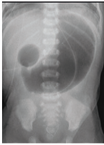
A 2-week-old boy has developed bilious vomiting. He was born via cesarean section at term. On physical exam, his pulse is 140, blood pressure is 80/50 mmHg, and respirations are 40/min. His abdomen appears distended and appears diffusely tender to palpation. Abdominal imaging is obtained (Figures A). Which of the following describes the mechanism that caused this child's disorder?
A 4-month-old girl is brought to the office by her parents because they noticed a mass protruding from her rectum and, she has been producing green colored emesis for the past 24 hours. Her parents noticed the mass when she had a bowel movement while changing her diaper. She strained to have this bowel movement 24 hours ago, shortly afterwards she had 3 episodes of greenish vomiting. She has a past medical history of failure to pass meconium for 2 days after birth. Her vital signs include: heart rate 190/min, respiratory rate 44/min, temperature 37.2°C (99.0°F), and blood pressure 80/50 mm Hg. On physical examination, the abdomen is distended. Examination of the anus reveals extrusion of the rectal mucosa through the external anal sphincter, and digital rectal examination produces an explosive expulsion of gas and stool. The abdominal radiograph shows bowel distention and absence of distal gas. What is the most likely cause?
A 3200-g (7.1-lb) female newborn is delivered at 38 weeks' gestation to a 24-year-old woman. The mother had regular prenatal visits throughout the pregnancy. The newborn's blood pressure is 53/35 mm Hg. Examination in the delivery room shows clitoromegaly and posterior labial fusion. One day later, serum studies show: Na+ 131 mEq/L K+ 5.4 mEq/L Cl− 102 mEq/L Urea nitrogen 15 mg/dL Creatinine 0.8 mg/dL Ultrasound of the abdomen and pelvis shows a normal uterus and ovaries. Further evaluation of the newborn is most likely to show which of the following findings?
A 4-year-old Caucasian male suffers from cyanosis and dyspnea relieved by squatting. Which of the following abnormalities is most likely present?
A 4-day-old male infant is brought to the physician because of respiratory distress and bluish discoloration of his lips and tongue. He was born at term and the antenatal period was uncomplicated. His temperature is 37.3°C (99.1°F), pulse is 170/min, respirations are 65/min, and blood pressure is 70/46 mm Hg. Pulse oximetry on room air shows an oxygen saturation of 82%. A grade 3/6 holosystolic murmur is heard over the left lower sternal border. A single S2 that does not split with respiration is present. Echocardiography shows defects in the interatrial and interventricular septae, as well as an imperforate muscular septum between the right atrium and right ventricle. Further evaluation of this patient is most likely to show which of the following?
A 7-month-old boy is brought to the physician for a well-child examination. He was born at 36 weeks' gestation and has been healthy since. He is at the 60th percentile for length and weight. Vital signs are within normal limits. The abdomen is soft and nontender. The external genitalia appear normal. Examination shows a single palpable testicle in the right hemiscrotum. The scrotum is nontender and not enlarged. There is a palpable mass in the left inguinal canal. Which of the following is the most appropriate next best step in management?
Practice by Chapter
Neural tube defects
Practice Questions
Congenital heart defects
Practice Questions
Gastrointestinal malformations
Practice Questions
Genitourinary anomalies
Practice Questions
Craniofacial anomalies
Practice Questions
Skeletal dysplasias
Practice Questions
Chromosomal disorders
Practice Questions
Teratogenic exposures
Practice Questions
Multiple malformation syndromes
Practice Questions
Prenatal diagnosis of congenital defects
Practice Questions
Surgical management timing
Practice Questions
Long-term outcomes and follow-up
Practice Questions
Preventive strategies
Practice Questions
Want unlimited practice?
Get full access to all questions, explanations, and performance tracking.
Scan to download app