
Common pediatric cancers — MCQs

On this page
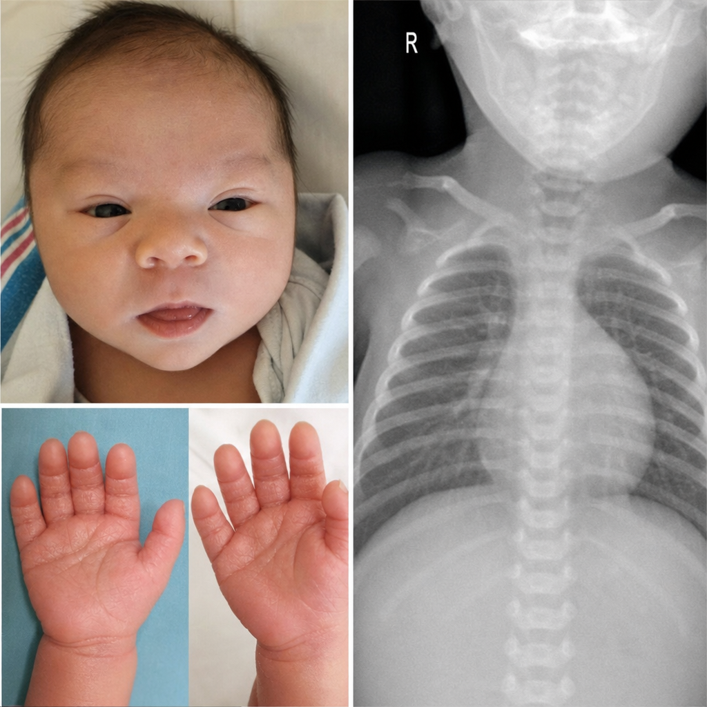
A newborn is delivered at 38 weeks gestation to a 28-year-old mother via spontaneous vaginal delivery. On examination in the delivery room, the infant has upslanting palpebral fissures, a flat nasal bridge, a single palmar crease bilaterally, and marked hypotonia. A harsh holosystolic murmur is heard at the left lower sternal border. The pediatric cardiology team is notified. Which of the following chromosomal findings is most consistent with this clinical presentation?
A 4-year-old boy with Down syndrome presents with fatigue and recurrent infections. CBC shows WBC 150,000/μL with 90% myeloblasts, hemoglobin 6.5 g/dL, platelets 15,000/μL. Flow cytometry confirms acute myeloid leukemia with megakaryoblastic features (AMKL). The parents are concerned about treatment intensity given their child's baseline developmental delays and increased treatment-related toxicity risk in Down syndrome. Evaluate the treatment approach considering the unique biology and competing risks.
A 15-year-old boy presents with right distal femur pain and a palpable mass. X-ray shows a mixed lytic-sclerotic lesion with periosteal elevation creating a Codman triangle and sunburst pattern. Biopsy confirms osteosarcoma. Staging shows pulmonary micrometastases. Alkaline phosphatase is markedly elevated. The family requests consideration of alternative therapies and limb salvage options. Synthesize the treatment plan addressing oncologic outcomes and functional preservation.
A 2-year-old girl presents with a large abdominal mass, aniridia, and developmental delay. Family history reveals a sibling who died of Wilms tumor at age 3. Genetic testing shows a germline WT1 mutation. Ultrasound reveals bilateral renal masses. The parents are concerned about treatment options that preserve renal function. Evaluate the optimal management strategy considering long-term outcomes.
A 6-year-old girl presents with acute onset left leg pain and refusal to bear weight. X-ray shows a lytic lesion with periosteal reaction in the femoral diaphysis demonstrating an 'onion-skin' pattern. MRI reveals a large soft tissue mass. Biopsy shows small round blue cells that are CD99 positive. Molecular studies show EWSR1-FLI1 fusion. Staging shows no metastases. Analyze the pathophysiology and treatment rationale.
Practice by Chapter
Leukemias in children
Practice Questions
Lymphomas in pediatric population
Practice Questions
Brain tumors in children
Practice Questions
Neuroblastoma
Practice Questions
Wilms tumor
Practice Questions
Rhabdomyosarcoma
Practice Questions
Retinoblastoma
Practice Questions
Ewing sarcoma
Practice Questions
Osteosarcoma
Practice Questions
Cancer predisposition syndromes
Practice Questions
Diagnostic approach to pediatric malignancies
Practice Questions
Pediatric oncology treatment principles
Practice Questions
Long-term survivor follow-up
Practice Questions
Want unlimited practice?
Get full access to all questions, explanations, and performance tracking.
Scan to download app