
Neoplasia — MCQs

On this page
A 14-year-old boy presents to his pediatrician with a 5-day history of abdominal pain and bloody stool. He denies having a fever and says that he has not experienced any other symptoms associated with the abdominal pain. He has no past medical history and does not take any medications or supplements. His family history is significant for a grandfather who developed Alzheimer disease at age 80 and a cousin who died at age 21 from colon cancer. Physical exam is unremarkable. Based on clinical suspicion a colonoscopy is obtained showing hundreds of small polyps in the colon. A mutation of a gene on which of the following chromosomes is most likely responsible for this patient's symptoms?
A 72-year-old and his caregiver present for a follow-up after a transthoracic needle biopsy of one of the large lesions in his chest was reported as non-small cell carcinoma of the lung. Previously, a chest CT revealed numerous nodules in the lungs bilaterally. The chest CT was ordered after the patient experienced a persistent cough with hemoptysis and a history of multiple episodes of pneumonia over the past year. The patient has a history of dementia and is a poor historian. The caregiver states that the patient has no history of smoking and that he was a lawyer before he retired, 10 years ago. The caregiver can only provide a limited medical history, but states that the patient sees another doctor “to monitor his prostate”. Which of the following is true regarding the pathogenesis of the nodules seen in this patient?
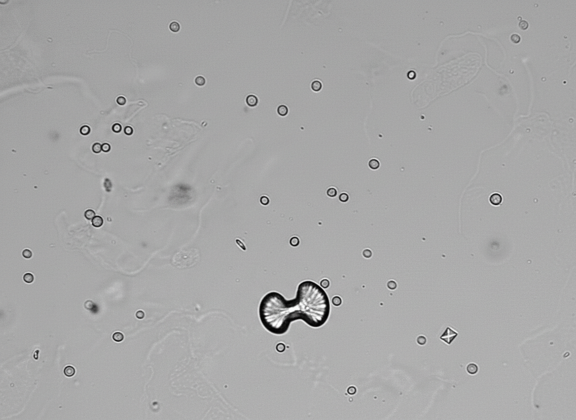
A 34-year-old patient with a history of anxiety, chronic constipation, chronic headaches, and chronic hypertension presents to the emergency room with severe right flank pain radiating to his scrotum. A urinalysis with stone analysis is performed and the results are shown in figure A. Prior to discharge, it is noted that the patients BP is still 170/110 mmHg. Furthermore, his calcium and PTH levels were both found to be increased. Which of the following representative histology slides of thyroid tissue represents a potential complication of the patients condition?
An 82-year-old man is brought to the emergency department after he was found down by his daughter. On presentation, he is alert and oriented with no obvious signs of trauma. He says that he felt lightheaded shortly before passing out and that he has been feeling extremely fatigued over the last few weeks. He has a known diagnosis of colorectal adenocarcinoma and had it surgically removed 2 months ago; however, recently he has been feeling increasingly short of breath. He has a 60-pack-year smoking history and drinks 2-3 beers a night. He worked as an insulation technician and shipyard laborer for 40 years prior to retiring at age 65. Radiographs reveal approximately a dozen new nodules scattered throughout his lungs bilaterally. Biopsy of these lesions would most likely reveal which of the following?
A 22-year-old man comes to the physician because of headaches and blurry vision for the past 6 months. He also reports frequent episodes of vomiting over the last month. His father has died of renal cell carcinoma at the age of 37 years. Examination shows 20/40 vision bilaterally. Fundoscopic examination shows bilateral optic disc swelling and growth of capillary vessels in the temporal peripheral retina. An MRI of the brain shows an infratentorial mass. The patient undergoes surgical resection of the mass. A photomicrograph of the resected specimen is shown. Which of the following is the most likely diagnosis?
A 16-year-old boy is brought to the physician because of a lesion that has been growing on his jaw over the past several months. He recently immigrated to the USA from Kenya with his family. Physical examination shows a 3-cm solid mass located above the left mandible. There is cervical lymphadenopathy. Biopsy of the mass shows sheets of lymphocytes and interspersed reactive histiocytes with abundant, clear cytoplasm and phagocytosed debris. Which of the following mechanisms is most likely directly responsible for the malignant transformation of this patient's cells?
A 39-year-old African American woman is admitted to the hospital following a seizure with a severe post-ictal headache. She was diagnosed with breast cancer 1 year ago when she presented with a hard, rock-like, immobile mass with irregular borders accompanied by changes in the breast skin, including erythema and dimpling. She had ipsilateral mobile axillary lymphadenopathy at that time. A biopsy confirmed the diagnosis of stage 2B invasive breast cancer. Her mother died at 42 years of age due to the same type of breast cancer. A CT scan done during this admission reveals multiple metastatic lesions in the brain and liver, along with the involvement of supra- and infra-clavicular lymph nodes. Which of the following molecular profile most likely characterizes this patient?
A 53-year-old farmer presents to the clinic for evaluation of a pigmented lesion on his arm. He states that he first noticed the lesion last year, but he believes that it has been slowly growing in size. He otherwise does not have any complaints and is generally healthy. Which of the following findings on physical exam would suggest a malignant diagnosis?
A 40-year-old man presents with an episode of rectal bleeding. He is concerned because his mother died of colorectal cancer at 50 years of age. He has no further information about his family history. Physical examination and digital rectal examination are normal. He undergoes a colonoscopy and is found to have innumerable adenomas in the left side of the colon ranging in size from 4–15 mm. Which of the following is the most likely underlying mechanism of this patient illness?
A 55-year-old woman comes to the physician because of a 4-month history of a painless lump on her neck. Examination shows a hard nodule on the left side of her neck. A fine-needle aspiration biopsy shows well-differentiated cuboidal cells arranged spherically around colloid. She undergoes thyroidectomy. Histopathological examination of the surgical specimen shows invasion of the thyroid capsule and blood vessels. Which of the following cellular events is most likely involved in the pathogenesis of this patient's condition?
Practice by Chapter
Characteristics of benign vs malignant tumors
Practice Questions
Nomenclature of neoplasms
Practice Questions
Carcinogenesis models
Practice Questions
Oncogenes and proto-oncogenes
Practice Questions
Tumor suppressor genes
Practice Questions
DNA repair genes and cancer
Practice Questions
Epigenetic mechanisms in cancer
Practice Questions
Apoptosis and cancer
Practice Questions
Tumor angiogenesis
Practice Questions
Tumor invasion and metastasis
Practice Questions
Carcinogenic agents
Practice Questions
Paraneoplastic syndromes
Practice Questions
Tumor immunology
Practice Questions
Want unlimited practice?
Get full access to all questions, explanations, and performance tracking.
Scan to download app