
Neoplasia — MCQs

On this page
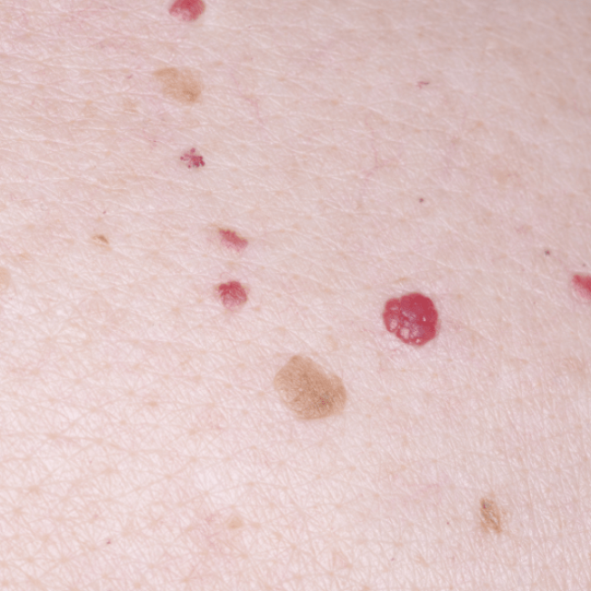
A 51-year-old woman presents to the dermatologist with concern for a new skin lesion (Image A). You note two similar lesions on her back. Which of the following is a true statement about these lesions?
A 70-year-old man comes to the physician because of a painless skin lesion on his neck for the past 5 months. The lesion has gradually become darker in color and is often pruritic. He has a similar lesion on the back. He is a retired landscaper. He has smoked half a pack of cigarettes daily for 45 years. Physical examination shows a 0.9-cm hyperpigmented papule on the neck with a greasy, wax-like, and stuck-on appearance. Histopathologic examination is most likely to show which of the following?
A 2-year-old boy is brought to the physician with complaints of gingival growth in the lower jaw with associated pain for the past few weeks. He has no history of trauma or any other significant medical conditions. His temperature is 37.0°C (98.6°F), pulse is 92/min, and respiratory rate is 24/min. On extraoral examination, a swelling of 4 cm x 2 cm is present on the left lower jaw. On intraoral examination, a diffuse erythematous swelling covered with necrotic slough is present on the gingiva. Computed tomography (CT) scan of the head shows multiple soft tissue density lesions involving mandibular, maxillary, left occipital, and temporal regions. Which of the following findings, if present, would be the most specific indicator of the disease in this patient?
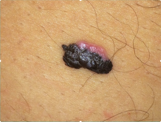
A 59-year-old man comes to the physician for evaluation of a progressively enlarging, 8-mm skin lesion on the right shoulder that developed 1 month ago. The patient has a light-skinned complexion and has had several dysplastic nevi removed in the past. A photograph of the lesion is shown. The lesion is most likely derived from cells that are also the embryological origin of which of the following tumors?
A 45-year-old man comes to the physician for the evaluation of difficulty swallowing that has worsened over the past year. He also reports some hoarseness and generalized bone, muscle, and joint pain. During the past six months, he has had progressive constipation and two episodes of kidney stones. He also reports recurrent episodes of throbbing headaches, diaphoresis, and palpitations. He does not smoke or drink alcohol. He takes no medications. His vital signs are within normal limits. Physical examination and an ECG show no abnormalities. Laboratory studies show calcium concentration of 12 mg/dL, phosphorus concentration of 2 mg/dL, alkaline phosphatase concentration of 100 U/L, and calcitonin concentration of 11 pg/mL (N < 8.8). Ultrasonography of the neck shows hypoechoic thyroid lesions with irregular margins and microcalcifications. Which of the following is the most likely underlying cause of this patient's condition?
A previously healthy 68-year-old woman is brought to the emergency department because of a 3-day history of nausea, anorexia, polyuria, and confusion. Her only medication is acetaminophen, which she takes daily for back pain that started 6 weeks ago. Physical examination shows conjunctival pallor. She is oriented to person but not to time or place. Laboratory studies show a hemoglobin concentration of 9.3 g/dL, a serum calcium concentration of 13.8 mg/dL, and a serum creatinine concentration of 2.1 mg/dL. Her erythrocyte sedimentation rate is 65 mm/h. Which of the following is the most likely underlying cause of this patient's condition?
A 58-year-old woman presents to a physician with a painless swelling behind her right ear, which she noticed 1 month ago. She has no other complaint nor does she have any specific medical condition. On physical examination, her vital signs are stable. An examination of the right post-auricular area shows enlarged lymph nodes, which are non-tender and rubbery in consistency, with normal overlying skin. A detailed general examination reveals the presence of one enlarged axillary lymph node on the left side with similar features. Complete blood counts are within normal limits but atypical lymphocytes are present on the peripheral blood smear. The patient’s serum lactate dehydrogenase level is slightly elevated. Excisional biopsy of the lymph node is performed and histopathological examination of the tissue yields a diagnosis of follicular lymphoma. Further cytogenetic studies reveal that the condition is associated with overexpression of the BCL-2 gene. Which of the following cytogenetic abnormalities is most likely to be present?
A 33-year-old woman comes to the physician 1 week after noticing a lump in her right breast. Fifteen years ago, she was diagnosed with osteosarcoma of her left distal femur. Her father died of an adrenocortical carcinoma at the age of 41 years. Examination shows a 2-cm, firm, immobile mass in the lower outer quadrant of the right breast. A core needle biopsy of the mass shows adenocarcinoma. Genetic analysis in this patient is most likely to show a defect in which of the following genes?
A 30-year-old boxer seeks evaluation by his physician after he noticed swelling at the angle of his jaw a few days ago. He recalls a recent boxing match when he was punched in his face. He says that his jaw is very painful. On examination, a firm mass is palpated, measuring 4 x 4 cm. An ultrasound was performed, which shows a thin, encapsulated, well-circumscribed, predominantly solid mass with occasional cystic areas. The mass is surgically excised, after which he develops a hoarse voice for a few days, but recovers within 1 week. The histopathologic evaluation of the surgical specimen reports a pseudocapsule with a hypocellular stromal component consisting of a myxoid background and cartilage arranged in clusters and a hypercellular epithelial component with cells arranged in sheets and trabeculae. From which of the following structures did the mass most likely arise?
A 62-year-old woman presents to her primary care physician because of fever, fatigue, and shortness of breath. She has noticed that she has a number of bruises, but she attributes this to a hike she went on 1 week ago. She has diabetes and hypertension well controlled on medication and previously had an abdominal surgery but doesn’t remember why. On physical exam, she has some lumps in her neck and a palpable liver edge. Peripheral blood smear shows white blood cells with peroxidase positive eosinophilic cytoplasmic inclusions. The abnormal protein most likely seen in this disease normally has which of the following functions?
Practice by Chapter
Characteristics of benign vs malignant tumors
Practice Questions
Nomenclature of neoplasms
Practice Questions
Carcinogenesis models
Practice Questions
Oncogenes and proto-oncogenes
Practice Questions
Tumor suppressor genes
Practice Questions
DNA repair genes and cancer
Practice Questions
Epigenetic mechanisms in cancer
Practice Questions
Apoptosis and cancer
Practice Questions
Tumor angiogenesis
Practice Questions
Tumor invasion and metastasis
Practice Questions
Carcinogenic agents
Practice Questions
Paraneoplastic syndromes
Practice Questions
Tumor immunology
Practice Questions
Want unlimited practice?
Get full access to all questions, explanations, and performance tracking.
Scan to download app