
Inflammation — MCQs

On this page
A 28-year-old man is referred to the dermatologist for 2 months of increasing appearance of multiple smooth, circular patches of complete hair loss on his scalp. He says that the patches have associated pruritus and a burning sensation, and are not improving with the over-the-counter products recommended by his hair stylist. He denies pulling his hair intentionally. Physical examination reveals no epidermal inflammation or erythema, and no fluorescence is detected under Wood’s lamp. A punch biopsy shows a peribulbar lymphocytic inflammatory infiltrate surrounding anagen follicles, resembling a swarm of bees. Which of the following is the most likely diagnosis in this patient?
A 15-year-old girl is brought to the physician by her mother for a 2-day history of abdominal pain, nausea, vomiting, diarrhea, and decreased appetite. Her last menstrual period was 3 weeks ago. Her temperature is 37.6°C (99.7°F). Abdominal examination shows tenderness to palpation with guarding in the right lower quadrant. Laboratory studies show a leukocyte count of 12,600/mm3. Which of the following is the most likely underlying cause of this patient's condition?
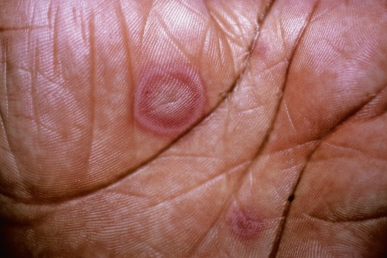
A 19-year-old man presents with painful oral ulcers and rash. He says that his symptoms started 1 week ago with a low-grade fever, malaise, and cough. Then, 3 days ago, he noted small painful red bumps on his hands and feet, which quickly worsened and spread to involve his extremities and upper torso. At the same time, multiple painful oral ulcers appeared, which have not improved. He denies any trouble breathing, pruritus, hemoptysis, hematochezia, or similar symptoms in the past. Past medical history is significant for a recent methicillin-resistant staphylococcus aureus (MRSA) skin infection 2 weeks ago secondary to a laceration on his left leg for which he has been taking trimethoprim-sulfamethoxazole. No other current medications. The patient is afebrile, and his vital signs are within normal limits. Physical examination reveals multiple raised, erythematous, circular papules averaging 1–2 cm in diameter with a central bulla, as shown in the exhibit (see image below). The cutaneous lesions occupy < 10% of his total body surface area (BSA). Nikolsky sign is negative. Multiple mucosal erosions are noted in the oral cavity. Generalized lymphadenopathy is present. A well-healing laceration is present on the left leg with no evidence of drainage or fluctuance. A cutaneous punch biopsy of one of the lesions is performed. Which of the following histopathologic features would most likely be found on this patient's biopsy?
A 34-year-old woman comes to the physician because of a 6-week history of fever and productive cough with blood-tinged sputum. She has also had a 4-kg (8.8-lb) weight loss during the same time period. Examination shows enlarged cervical lymph nodes. An x-ray of the chest shows a 2.5-cm pulmonary nodule in the right upper lobe. A biopsy specimen of the lung nodule shows caseating granulomas with surrounding multinucleated giant cells. Which of the following is the most likely underlying cause of this patient's pulmonary nodule?
A 36-year-old man is brought to the emergency department by his wife 20 minutes after having a seizure. Over the past 3 days, he has had a fever and worsening headaches. This morning, his wife noticed that he was irritable and demonstrated strange behavior; he put the back of his fork, the salt shaker, and the lid of the coffee can into his mouth. He has no history of serious illness and takes no medications. His temperature is 39°C (102.2°F), pulse is 88/min, and blood pressure is 118/76 mm Hg. Neurologic examination shows diffuse hyperreflexia and an extensor response to the plantar reflex on the right. A T2-weighted MRI of the brain shows edema and areas of hemorrhage in the left temporal lobe. Which of the following is most likely the primary mechanism of the development of edema in this patient?
A 3-year-old male is brought by his mother to the pediatrician because she is concerned about a lump in his neck. She reports that the child was recently ill with a cough, nasal congestion, and rhinorrhea. She also noticed that a small red lump developed on the patient’s neck while he was sick. Although his cough and congestion subsided after a few days, the neck lump has persisted. The child has no notable past medical history. He was born at 39 weeks gestation and is in the 55th percentiles for both height and weight. His temperature is 98.6°F (37°C), blood pressure is 105/65 mmHg, pulse is 90/min, and respirations are 18/min. Physical examination reveals a small, soft, rounded mass at the midline of the neck inferior to the hyoid bone. The mass is warm and tender to palpation. It moves superiorly when the patient drinks water. Histologic examination of this lesion would most likely reveal which of the following?
A 50-year-old female presents to her physician with vesicles and tense blisters across her chest, arms, and the back of her shoulders. Physical examination reveals that blistering is not present in her oral mucosa, and the epidermis does not separate upon light stroking of the skin. The patient most likely suffers from a hypersensitivity reaction located:
A 6-year-old boy is brought to the emergency department due to a severe infection. Laboratory work shows leukocytosis of 60 × 109/L with marked left shift, but no blast cells. The patient is febrile and dehydrated. The physician believes that this is a severe reaction to the infection and orders a leukocyte alkaline phosphatase (LAP) stain on a peripheral smear. The LAP score is elevated. Which of the following statements best describes an additional characteristic of the condition this child is suffering from?
A 40-year-old man presents with a swollen left big toe that started this morning. The patient states that he attended a party last night and drank 4 glasses of whiskey. He denies any trauma to the foot. The patient has a history of similar episodes in the past that were related to alcohol use. His symptoms were previously relieved with ibuprofen. However, the pain persisted despite treatment with the medication. Physical examination reveals a tender and erythematous, swollen left 1st metatarsophalangeal joint. Which of the following events most likely contributed to his condition?
A 42-year-old woman with well-controlled HIV on antiretroviral therapy comes to the physician because of a 2-week history of a painless lesion on her right calf. Many years ago, she had a maculopapular rash over her trunk, palms, and soles that resolved spontaneously. Physical examination shows a 4-cm firm, non-tender, indurated ulcer with a moist, dark base and rolled edges. There is a similar lesion at the anus. Results of rapid plasma reagin testing are positive. Which of the following findings is most likely on microscopic examination of these lesions?
Practice by Chapter
Acute inflammation mechanisms
Practice Questions
Vascular changes in inflammation
Practice Questions
Chemical mediators of inflammation
Practice Questions
Cellular components of inflammation
Practice Questions
Resolution of acute inflammation
Practice Questions
Chronic inflammation
Practice Questions
Granulomatous inflammation
Practice Questions
Systemic effects of inflammation
Practice Questions
Patterns of inflammatory response
Practice Questions
Inflammatory markers in laboratory testing
Practice Questions
Anti-inflammatory therapies
Practice Questions
Wound healing and repair
Practice Questions
Abnormalities in wound healing
Practice Questions
Want unlimited practice?
Get full access to all questions, explanations, and performance tracking.
Scan to download app