
Peritonitis — MCQs

A 40-year-old man presents with acute abdominal pain. Past medical history is significant for hepatitis C, complicated by multiple recent visits with associated ascites. His temperature is 38.3°C (100.9°F), heart rate is 115/min, blood pressure is 88/48 mm Hg, and respiratory rate is 16/min. On physical examination, the patient is alert and in moderate discomfort. Cardiopulmonary examination is unremarkable. Abdominal examination reveals distant bowel sounds on auscultation. There is also mild diffuse abdominal tenderness to palpation with guarding present. The remainder of the physical examination is unremarkable. A paracentesis is performed. Laboratory results are significant for the following: Leukocyte count 11,630/µL (with 94% neutrophils) Platelets 24,000/µL Hematocrit 29% Ascitic fluid analysis: Cell count 658 PMNs/µL Total protein 1.2 g/dL Glucose 24 mg/dL Gram stain Gram-negative rods Culture Culture yields growth of E. coli Which of the following is the next, best step in the management of this patient?
A 49-year-old woman with a history of hepatitis C cirrhosis complicated by esophageal varices, ascites, and hepatic encephalopathy presents with 1 week of increasing abdominal discomfort. Currently, she takes lactulose, rifaximin, furosemide, and spironolactone. On physical examination, she has mild asterixis, generalized jaundice, and a distended abdomen with positive fluid wave. Diagnostic paracentesis yields a WBC count of 1196/uL with 85% neutrophils. Which of the following is the most appropriate treatment?
A 15-year-old boy presents with his father to the urgent care with 5 days of frequent diarrhea, occasionally with streaks of blood mixed in. Stool cultures are pending, but preliminary stool samples demonstrate fecal leukocytes and erythrocytes. His vital signs are as follows: blood pressure is 126/83 mm Hg, heart rate is 97/min, and respiratory rate is 15/min. He is started on outpatient therapy for presumed Shigella infection. Which of the following is the most appropriate therapy?
A 58-year-old man with liver cirrhosis presents to his primary care physician complaining of increased abdominal girth and early satiety. He drinks 2–4 glasses of wine with dinner and recalls having had abnormal liver enzymes in the past. Vital signs include a temperature of 37.1°C (98.7°F), blood pressure of 110/70 mm Hg, and a pulse of 75/min. Physical examination reveals telangiectasias, mild splenomegaly, palpable firm liver, and shifting dullness. Liver function is shown: Total bilirubin 3 mg/dL Aspartate aminotransferase (AST) 150 U/L Alanine aminotransferase (ALT) 70 U/L Total albumin 2.5 g/dL Abdominal ultrasonography confirms the presence of ascites. Diagnostic paracentesis is performed and its results are shown: Polymorphonuclear cell count 10 cells/mm Ascitic protein 1 g/dL Which of the following best represents the mechanism of ascites in this patient?
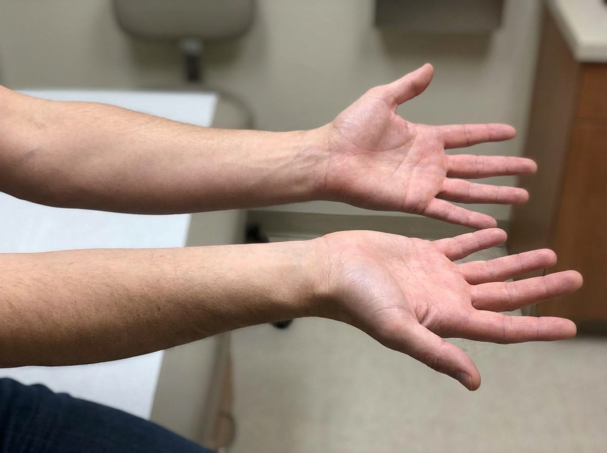
A 56-year-old male with a history of hepatitis C cirrhosis status post TIPS procedure is brought in by his wife to the emergency department because he has been acting disoriented, slurring his speech, and sleeping throughout the day. On arrival the patient is afebrile and his vital signs are pulse is 87/min, blood pressure is 137/93 mmHg, and respirations are 12/min with shallow breaths. Examination reveals a jaundiced male who appears older than stated age. Abdominal exam is positive for a fluid wave and shifting dullness to percussion. You note enlarged breasts, decreased facial hair, 3+ patellar reflexes bilaterally, and the following in the upper extremity (Image A). Paracentesis reveals ascitic fluid with neutrophil counts of < 100 cells/mcL. Serum creatinine is 1.0 and BUN is 15. Which of the following is the next best step in management?
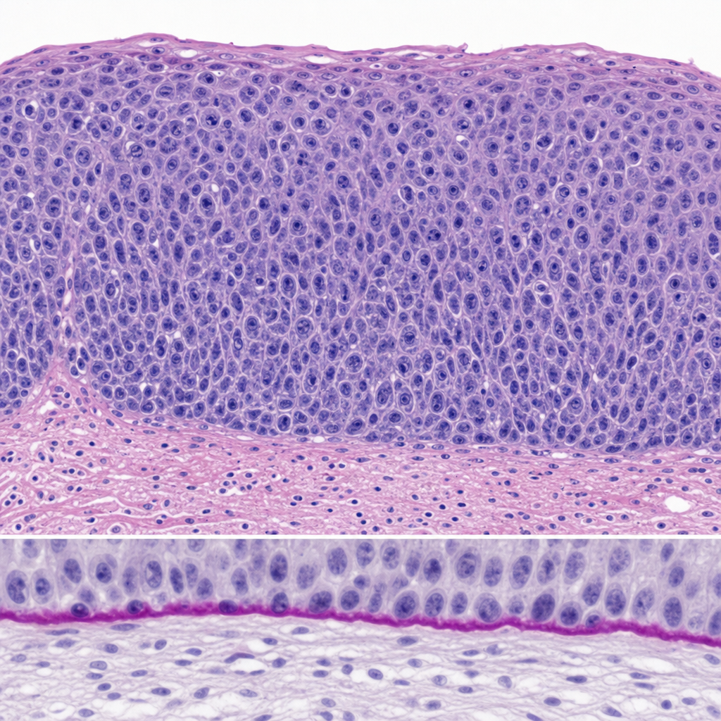
A 34-year-old woman undergoes excisional biopsy of a cervical lesion detected on colposcopy. The photomicrograph demonstrates full-thickness replacement of the squamous epithelium by atypical cells with high nuclear-to-cytoplasmic ratios, loss of polarity, and numerous mitotic figures extending to the surface. The basement membrane appears intact on PAS stain. Which of the following statements most accurately characterizes the biological behavior of this lesion?
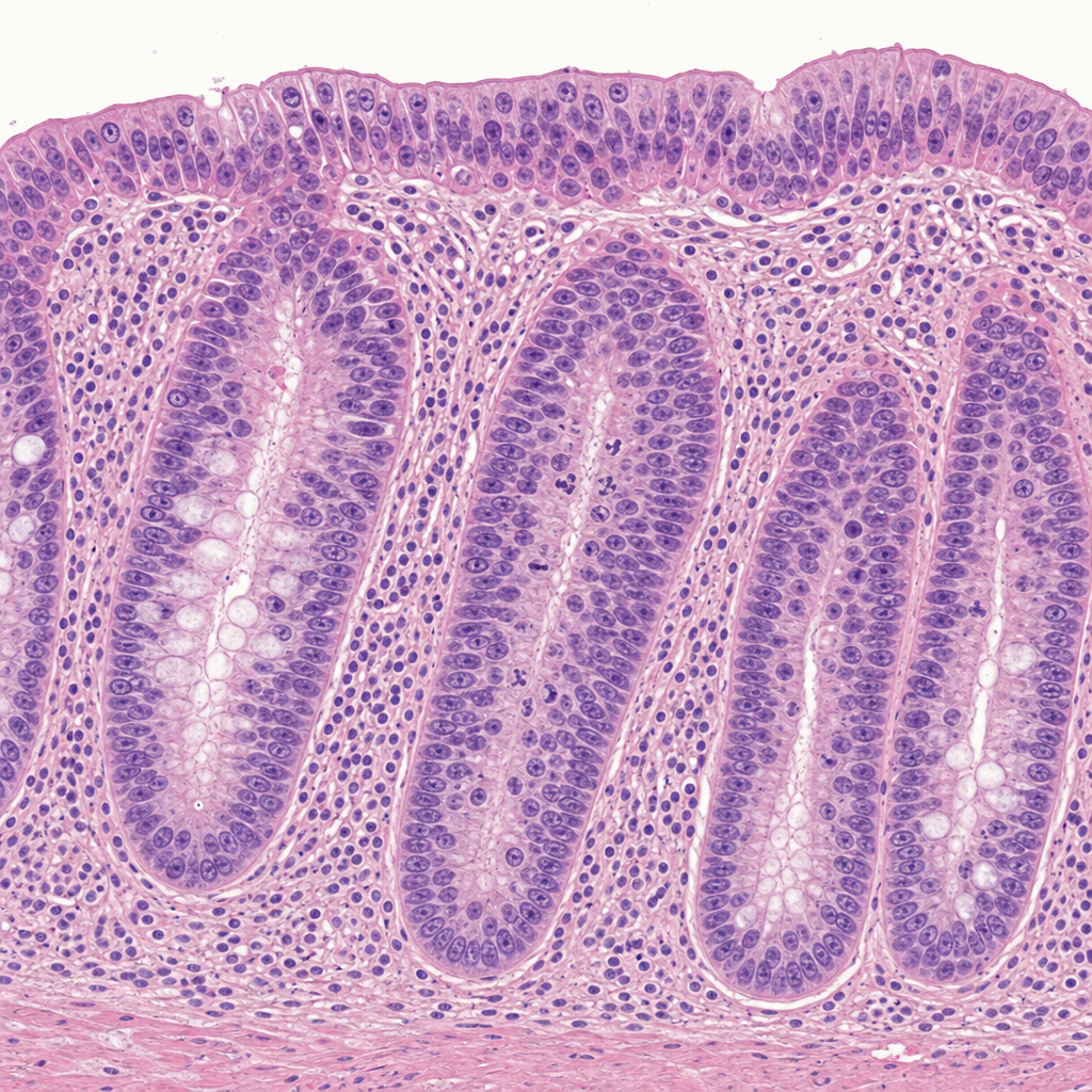
A 38-year-old woman with a history of ulcerative colitis for 12 years undergoes surveillance colonoscopy. A flat mucosal lesion is biopsied from the sigmoid colon. The photomicrograph shows colonic crypts with enlarged, hyperchromatic, stratified nuclei that extend to the luminal surface; nuclear polarity is partially lost; there is increased mitotic activity; and the basement membrane is intact with no extension of atypical cells into the lamina propria. Which of the following statements best characterizes the biological behavior of this lesion?
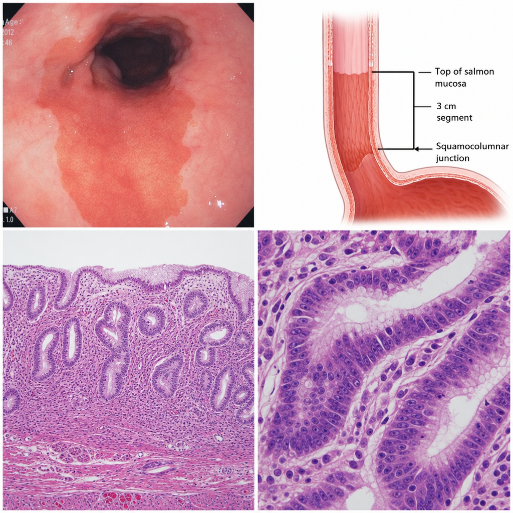
A 45-year-old woman with a history of chronic gastroesophageal reflux disease undergoes upper endoscopy. The gastroesophageal junction mucosa appears salmon-pink and velvety, extending 3 cm above the squamocolumnar junction. Biopsy is taken from this region. A subsequent biopsy taken one year later shows the same columnar epithelium with nuclear crowding, loss of polarity, and architectural complexity confined entirely above the basement membrane. Which of the following correctly describes the biological behavior of the change seen on the second biopsy?
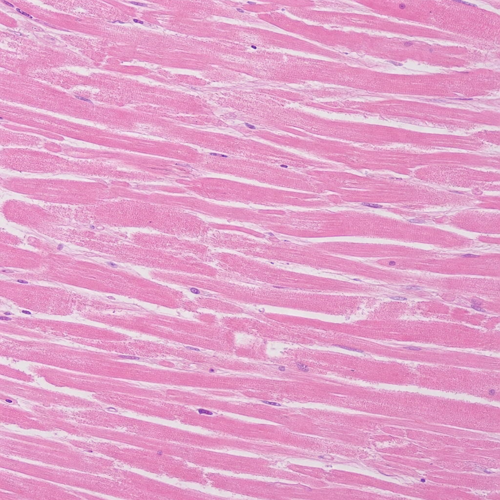
A 62-year-old man with a 40 pack-year smoking history dies suddenly. At autopsy, a large transmural infarct is identified in the left anterior descending artery territory. Histological sections from the center of the infarct are taken. The photomicrograph shows preserved cellular outlines with loss of nuclei and cytoplasmic eosinophilia, but no neutrophilic infiltrate is visible. No neutrophil margination is identified at the infarct periphery. Which of the following best estimates the age of this infarct, and which morphological feature most directly supports that estimate?
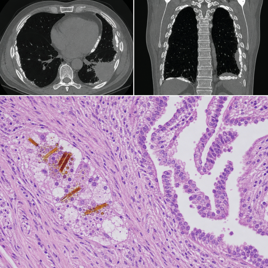
A 58-year-old man with a 40-pack-year smoking history presents with progressive dyspnea and a dry cough. Chest CT reveals bilateral pleural plaques and a pleural-based mass. A pleural biopsy is performed. The photomicrograph shows elongated, beaded, golden-brown structures within macrophages in the lung parenchyma adjacent to the mass, surrounded by a biphasic proliferation of epithelioid and spindle cells arranged in a tubulopapillary pattern. Which of the following best describes the golden-brown structures seen in this biopsy?
Want unlimited practice?
Get full access to all questions, explanations, and performance tracking.
Scan to download app