
Laboratory techniques in pathology — MCQs

An investigator studying the molecular characteristics of various malignant cell lines collects tissue samples from several families with a known mutation in the TP53 tumor suppressor gene. Immunohistochemical testing performed on one of the cell samples stains positive for desmin. This sample was most likely obtained from which of the following neoplasms?
During an experiment, the immunophenotypes of different cells in a sample are determined. The cells are labeled with fluorescent antibodies specific to surface proteins, and a laser is then focused on the samples. The intensity of fluorescence created by the laser beam is then plotted on a scatter plot. The result shows most of the cells in the sample to be positive for CD8 surface protein. Which of the following cell types is most likely represented in this sample?
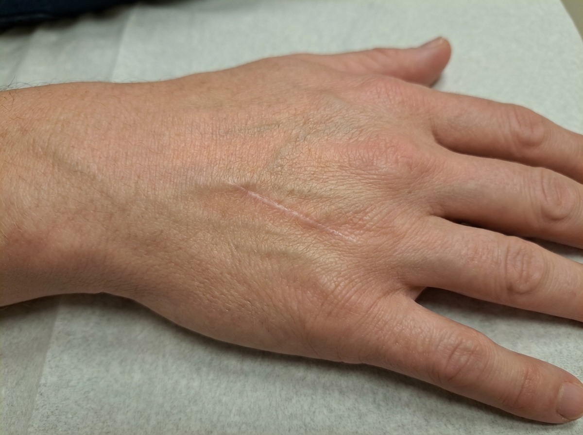
A 16-year-old boy presents to the emergency department after a skateboarding accident. He fell on a broken bottle and received a 4 cm wound on the dorsal aspect of his left hand. His vitals are stable and he was evaluated by the surgeon on call who determined that suturing was not required. After several weeks the wound has almost completely healed (see image). Which of the following is the correct description of this patient's wound before healing?
An investigator is processing a blood sample from a human subject. A reagent is added to the sample and the solution is heated to break the hydrogen bonds between complementary base pairs. This solution is then cooled to allow artificial DNA primers in the solution to attach to the separated strands of the sample DNA molecules. An enzyme derived from the thermophilic bacterium Thermus aquaticus is added and the solution is reheated. These steps are repeated multiple times until the aim of the test is achieved. The investigator most likely used which of the following laboratory procedures on the test sample?
A pathologist receives a patient sample for analysis. Cells in the sample are first labeled with fluorescent antibodies and then passed across a laser beam in a single file of particles. The light scatter and fluorescent intensity of the particles are plotted on a graph; this information is used to characterize the sample. This laboratory method would be most useful to establish the diagnosis of a patient with which of the following?
A 32-year-old man visits his family physician for 10 months of persistent left flank pain, weight loss, and fatigue. Also, he has had hematuria a couple of times in the last month. His mother was diagnosed and treated for a pheochromocytoma when she was 36 years old, and his father died at 45 years due to myocardial infarction. His personal medical history is not relevant. He does not smoke and used to be a varsity athlete in high school and university. Physical examination shows temporal wasting, pale mucous membranes and palms, a palpable mass in the left flank, and a varicocele that does not reduce upon recumbency. His family physician sends the patient to the emergency department for an abdominal computed tomography (CT) scan, which shows a complex left renal mass and a hemangioblastoma in T10. A biopsy of the renal mass is ordered by the oncology team, which demonstrates compact cells with prominent nucleoli, eosinophilic cytoplasm within a network of a small and thin-walled vasculature. What is the most likely type of tumor in this patient?
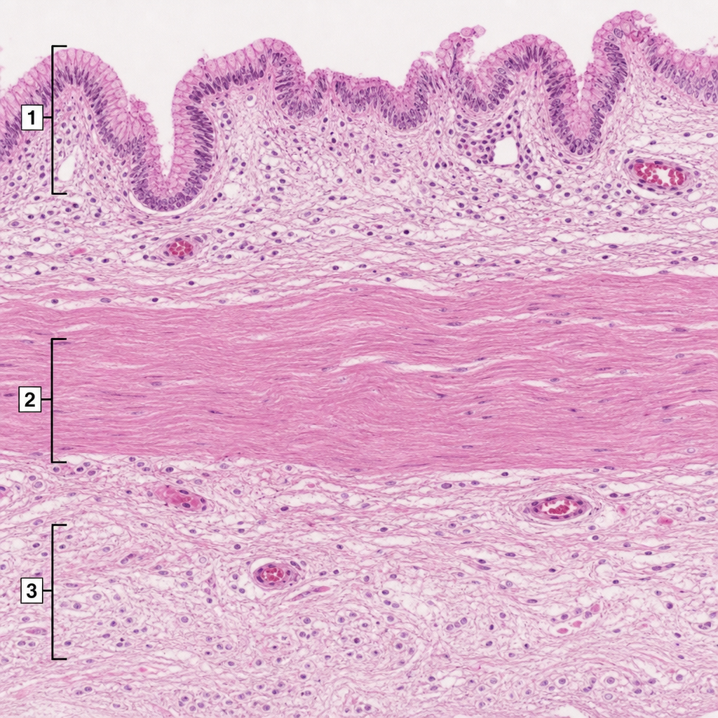
A 36-year-old man undergoes surgical intervention due to a right upper quadrant stab wound. His gallbladder was found to be lacerated and is removed. It is sent for histological evaluation. The pathologist examines the slide shown in the exhibit and identifies several structures numbered the image. Which of the following statements is correct?
A 72-year-old man goes to his primary care provider for a checkup after some blood work showed lymphocytosis 3 months ago. He says he has been feeling a bit more tired lately but doesn’t complain of any other symptoms. Past medical history is significant for hypertension and hyperlipidemia. He takes lisinopril, hydrochlorothiazide, and atorvastatin. Additionally, his right hip was replaced three years ago due to osteoarthritis. Family history is noncontributory. He drinks socially and does not smoke. Today, he has a heart rate of 95/min, respiratory rate of 17/min, blood pressure of 135/85 mm Hg, and temperature of 36.8°C (98.2°F). On physical exam, he looks well. His heartbeat has a regular rate and rhythm and lungs that are clear to auscultation bilaterally. Additionally, he has mild lymphadenopathy of his cervical lymph nodes. A complete blood count with differential shows the following: Leukocyte count 5,000/mm3 Red blood cell count 3.1 million/mm3 Hemoglobin 11.0 g/dL MCV 95 um3 MCH 29 pg/cell Platelet count 150,000/mm3 Neutrophils 40% Lymphocytes 40% Monocytes 5% A specimen is sent for flow cytometry that shows a population that is CD 5, 19, 20, 23 positive. Which of the following is the most likely diagnosis?
A 13-year-old boy is brought to the physician because of swelling around his eyes for the past 2 days. His mother also notes that his urine became gradually darker during this time. Three weeks ago, he was treated for bacterial tonsillitis. His temperature is 37.6°C (99.7°F), pulse is 79/min, and blood pressure is 158/87 mm Hg. Examination shows periorbital swelling. Laboratory studies show: Serum Urea nitrogen 9 mg/dL Creatinine 1.7 mg/dL Urine Protein 2+ RBC 12/hpf RBC casts numerous A renal biopsy would most likely show which of the following findings?
A 63-year-old man comes to the physician because of increasing generalized fatigue for 3 months. He is having more difficulty with keeping up with his activities of daily living and has lost 2.5 kg (5.5 lb) over the past month. He has hypertension and hyperlipidemia. He does not smoke and drinks two to three beers on weekends. His medications include lisinopril, hydrochlorothiazide, and atorvastatin. His temperature is 37.1°C (98.8°F), pulse is 85/min, respirations are 15/min, and blood pressure is 125/73 mm Hg. Examination shows pale conjunctivae. The remainder of the examination shows no abnormalities. His hematocrit is 27.3%, leukocyte count is 4500/mm3, and platelet count is 102,000/mm3. A peripheral blood smear shows numerous blast cells that stain positive for myeloperoxidase, CD33, and CD34. Which of the following is the most likely diagnosis?
Want unlimited practice?
Get full access to all questions, explanations, and performance tracking.
Scan to download app