
Disorders of pigmentation — MCQs

A 52-year-old Caucasian man presents to the clinic for evaluation of a mole on his back that he finds concerning. He states that his wife noticed the lesion and believes that it has been getting larger. On inspection, the lesion is 10 mm in diameter with irregular borders. A biopsy is performed. Pathology reveals abnormal melanocytes forming nests at the dermo-epidermal junction and discohesive cell growth into the epidermis. What is the most likely diagnosis?
A 19-year-old man presents to his primary care physician for evaluation before going off to college. Specifically, he wants to know how to stay healthy while living outside his home. Since childhood he has suffered severe sunburns even when he goes outside for a small period of time. He has also developed many freckles and rough-surfaced growths starting at the same age. Finally, his eyes are very sensitive and become irritated, bloodshot, and painful after being outside. A defect in a protein with which of the following functions is most likely responsible for this patient's symptoms?
A 53-year-old farmer presents to the clinic for evaluation of a pigmented lesion on his arm. He states that he first noticed the lesion last year, but he believes that it has been slowly growing in size. He otherwise does not have any complaints and is generally healthy. Which of the following findings on physical exam would suggest a malignant diagnosis?
A 27-year-old Caucasian female presents complaining of recent weight loss and weakness. She reports that she feels dizzy and lightheaded every morning when she gets out of bed, and often at work whenever she must rise from her desk. Physical exam reveals several areas of her skin including her elbows and knees are more pigmented than other areas. Which of the following would be consistent with the patient's disease?
A 52-year-old woman sees you in your office with a complaint of new-onset headaches over the past few weeks. On exam, you find a 2 x 2 cm dark, irregularly shaped, pigmented lesion on her back. She is concerned because her father recently passed away from skin cancer. What tissue type most directly gives rise to the lesion this patient is experiencing?
A 17-year-old Latin American woman with no significant past medical history or family history presents to her pediatrician with concerns about several long-standing skin lesions. She notes that she has had a light-colored rash on her chest and abdomen that has been present for the last 2 years. The blood pressure is 111/81 mm Hg, pulse is 82/min, respiratory rate is 16/min, and temperature is 37.3°C (99.1°F). Physical examination reveals numerous hypopigmented macules over her chest and abdomen. No lesions are seen on her palms or soles. When questioned, she states that these lesions do not tan like the rest of her skin when exposed to the sun. The remainder of her review of systems is negative. What is the most likely cause of these lesions?
A 28-year-old woman presents with progressive darkening of her face that started during pregnancy. The pigmentation is symmetrical and primarily affects her cheeks and forehead. It became more prominent with sun exposure. Examination reveals bilateral, symmetrical brown patches on her face. Which of the following is the most appropriate initial management?
An otherwise healthy 17-year-old girl comes to the physician because of multiple patches on her face, hands, abdomen, and feet that are lighter than the rest of her skin. The patches began to appear 3 years ago and have been gradually increasing in size since. There is no associated itchiness, redness, numbness, or pain. She emigrated from India 2 years ago. An image of the lesions on her face is shown. Which of the following is most likely involved in the pathogenesis of this patient's skin findings?

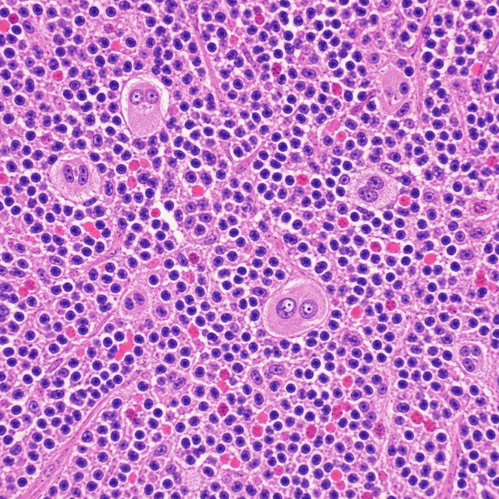
A 34-year-old woman presents with fatigue, night sweats, and a painless cervical lymph node mass for 3 months. Excisional biopsy of the lymph node is performed. The photomicrograph demonstrates a mixed cellular infiltrate with scattered large binucleated cells possessing prominent eosinophilic 'owl-eye' nucleoli, set against a background of lymphocytes, plasma cells, eosinophils, and fibroblasts. Immunohistochemical staining of the large cells is positive for CD15 and CD30 and negative for CD45. Which of the following best describes the cell of origin of the large binucleated cells seen in this lesion?
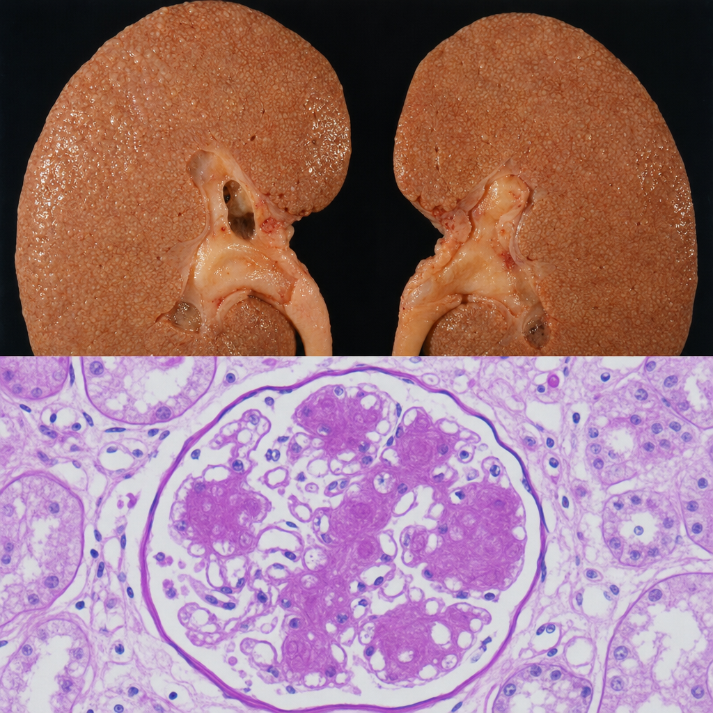
A 67-year-old man with a 20-year history of poorly controlled type 2 diabetes mellitus dies of a myocardial infarction. At autopsy, the kidneys are symmetrically enlarged with a granular cortical surface. PAS staining of the renal cortex shows PAS-positive ovoid deposits within the mesangium of glomeruli, compressing the adjacent capillary loops. Which of the following best describes the composition and pathogenetic mechanism of these deposits?
Want unlimited practice?
Get full access to all questions, explanations, and performance tracking.
Scan to download app