
Adnexal tumors — MCQs

A 7-year-old girl comes in to the emergency department with her mother for swelling of her left periorbital region. Yesterday morning she woke up with a painful, warm, soft lump on her left eyelid. Eye movement does not worsen the pain. Physical examination shows redness and swelling of the upper left eyelid, involving the hair follicles. Upon palpation, the swelling drains purulent fluid. Which of the following is the most likely diagnosis?
A 14-year-old boy presents as a new patient to your practice. While conducting your physical exam, you observe the findings depicted in Figures A and B. Which of the following additional findings would most likely be found in this patient?
A 16-year-old girl is brought to the physician by her mother because she has not had her menstrual period yet. At birth, she was found to have partial labial fusion and clitoromegaly. The mother reports that during the pregnancy she had noticed abnormal hair growth on her chin. The girl has severe acne. Three years ago, she broke her wrist after a minor trauma. Last year, she sustained a spinal compression fracture after lifting a box during a move. She currently takes oral isotretinoin and an oral contraceptive. The patient is at the 97th percentile for height and 50th percentile for weight. Physical examination shows numerous inflamed pustules on her face and upper back. Breast development is at Tanner stage I. The patient refuses to have a pelvic examination. A pelvic ultrasound shows ovaries with multiple cysts and a normal uterus. Which of the following is the most likely diagnosis?
A 27-year-old school teacher visits her doctor because of disfiguring skin lesions that started to appear in the past few days. The lesions are mostly located on her chest, shoulders, and back. They are 2–5 mm in diameter, droplike, erythematous papules with fine silver scales. Besides a sore throat and laryngitis requiring amoxicillin several weeks ago, she has no significant medical history. What is the most likely diagnosis?
A 9-year-old boy is brought to the physician for evaluation of 2 months of progressive clumsiness, falls, and increased urinary frequency. Physical examination shows bilateral temporal visual field loss. An MRI of the head shows a small calcified suprasellar mass. The patient undergoes surgery with complete removal of the mass. Pathological examination of the specimen shows a lobular tumor composed of cysts filled with oily, brownish-yellow fluid. This mass is most likely derived from which of the following structures?
A 55-year-old man with a history of sun exposure presents with a slowly growing, pearly nodule with telangiectasias on his nose. The lesion occasionally bleeds when traumatized. Biopsy shows basaloid cells arranged in palisading patterns. Which of the following mutations is most likely involved in the pathogenesis?
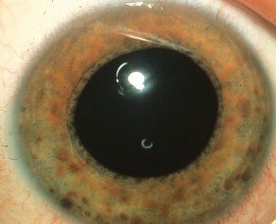
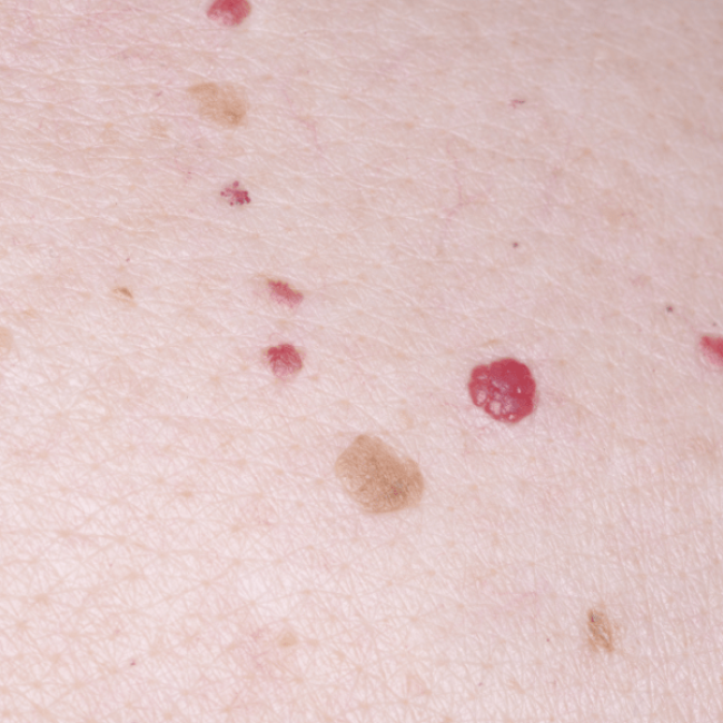
A 51-year-old woman presents to the dermatologist with concern for a new skin lesion (Image A). You note two similar lesions on her back. Which of the following is a true statement about these lesions?
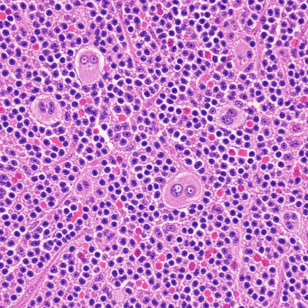
A 34-year-old woman presents with fatigue, night sweats, and a painless cervical lymph node mass for 3 months. Excisional biopsy of the lymph node is performed. The photomicrograph demonstrates a mixed cellular infiltrate with scattered large binucleated cells possessing prominent eosinophilic 'owl-eye' nucleoli, set against a background of lymphocytes, plasma cells, eosinophils, and fibroblasts. Immunohistochemical staining of the large cells is positive for CD15 and CD30 and negative for CD45. Which of the following best describes the cell of origin of the large binucleated cells seen in this lesion?
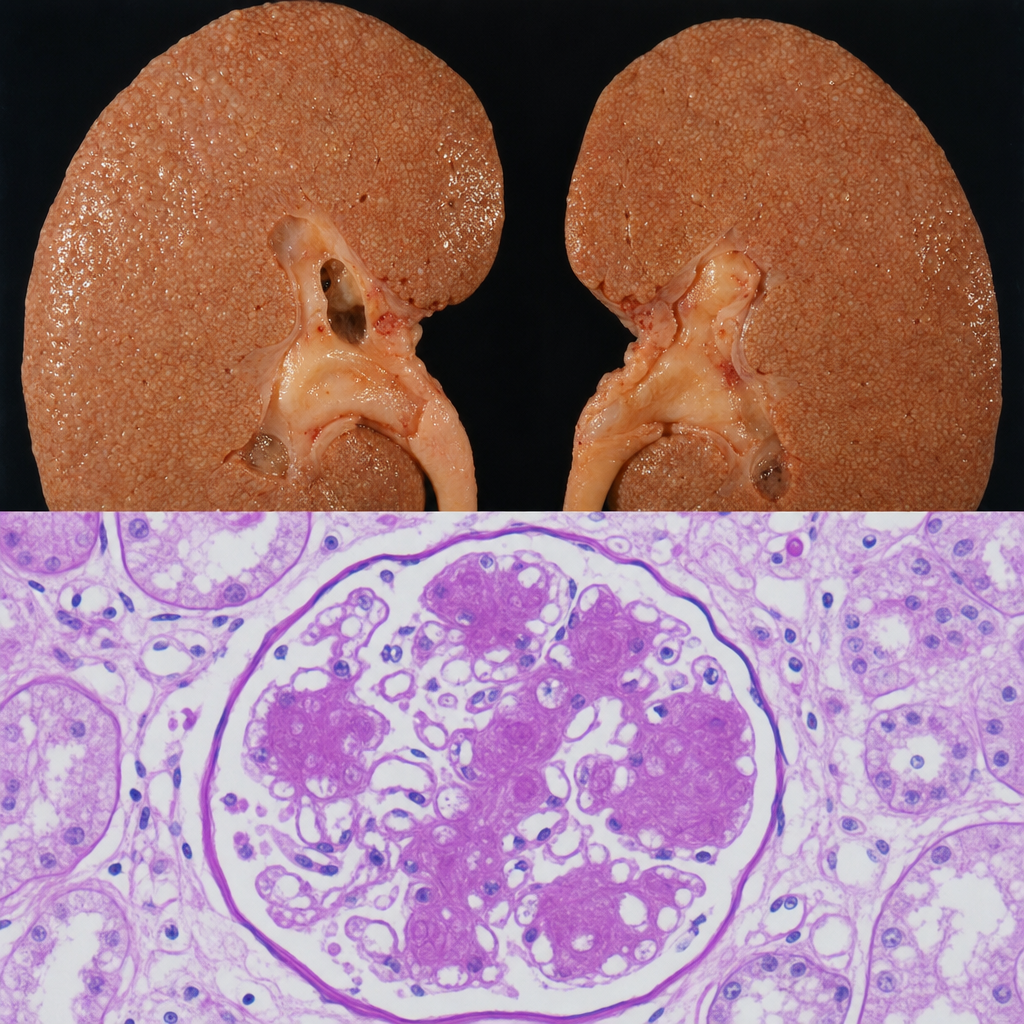
A 67-year-old man with a 20-year history of poorly controlled type 2 diabetes mellitus dies of a myocardial infarction. At autopsy, the kidneys are symmetrically enlarged with a granular cortical surface. PAS staining of the renal cortex shows PAS-positive ovoid deposits within the mesangium of glomeruli, compressing the adjacent capillary loops. Which of the following best describes the composition and pathogenetic mechanism of these deposits?
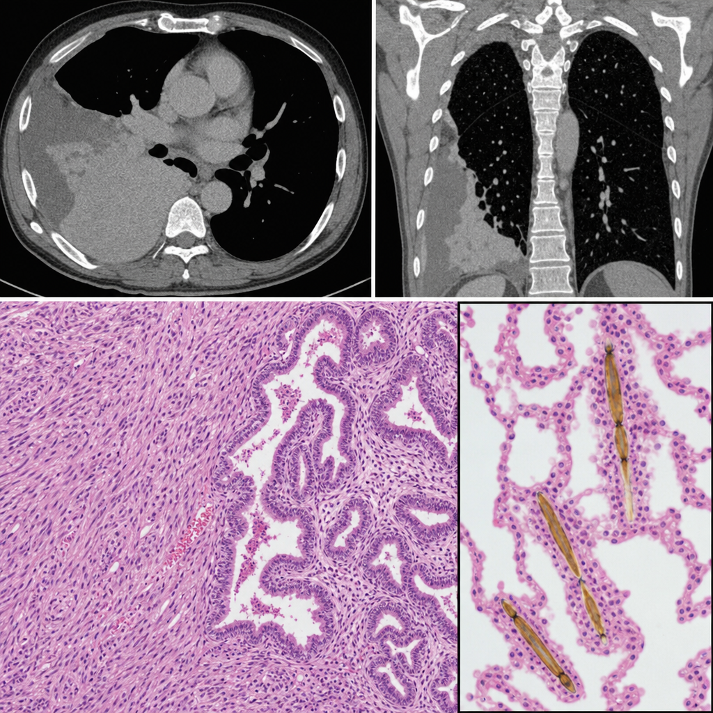
A 58-year-old man with a 40-pack-year smoking history presents with progressive dyspnea and a dry cough. Chest CT reveals bilateral pleural thickening and a right pleural mass. Thoracoscopic biopsy is performed. The photomicrograph shows a biphasic tumor with epithelioid cells forming tubulopapillary structures intermixed with a spindle-cell sarcomatoid component. Elongated, golden-brown, beaded structures with a translucent core are identified within the adjacent lung parenchyma, each coated with iron-protein complexes. Which of the following structures are most specifically represented by the coated inclusions seen in this specimen?
Want unlimited practice?
Get full access to all questions, explanations, and performance tracking.
Scan to download app