
Cell injury — MCQs

On this page
A 27-year-old man who recently immigrated to the United States with his family is diagnosed with an autosomal dominant disorder that causes anemia by extravascular hemolysis. The doctor explains that his red blood cells (RBCs) are spherical, which decreases their lifespan and explains that a splenectomy may be required in the future. Which of the following is most likely to be defective in this patient?
A 45-year-old man with a history of epilepsy comes to the physician for a follow-up examination. He has had trouble moving the right side of his body for 2 weeks. Three weeks ago he was admitted to the hospital for a generalized convulsive seizure. He was treated with intravenous lorazepam and phenytoin; the seizure activity resolved after 50 minutes on EEG monitoring. He was discharged 2 days later after no further epileptic activity occurred. Physical examination at discharge showed no abnormalities. He has had multiple hospitalizations for similar episodes over the past year. His only medication is lamotrigine, though he says that he sometimes forgets to take it. His temperature is 37°C (98.6°F), pulse is 70/min, and blood pressure is 130/80 mm Hg. Physical examination shows right-sided hemiparesis, right homonymous hemianopsia, and receptive aphasia. Which of the following is the most likely underlying cause of this patient's current symptoms?
An autopsy is performed on a 39-year-old man 5 days after he was found pulseless at his apartment by his neighbor. Examination of the brain shows liquefactive necrosis in the distribution of the right middle cerebral artery with surrounding edema. Immunophenotyping of a sample of the affected brain tissue shows numerous cells that express CD40 on their surface. On further histopathological evaluation, the morphology of these cells is not readily discernible with Nissl stain. These histological findings are most consistent with which of the following cell types?
As part of a clinical research study, microscopic analysis of tissues obtained from surgical specimens is performed. Some of these tissues have microscopic findings of an increase in the size of numerous cells within the tissue with an increase in the amount of cytoplasm, but the nuclei are uniform in size. Which of the following processes shows such microscopic findings?
A 59-year-old woman is admitted to the intensive care unit after surgery following a motor vehicle collision. She has received a total of four units of packed red blood cells. Physical examination shows dry mucous membranes and flat neck veins. Serum studies show a creatinine of 2.1 mg/dL and urine microscopy shows granular, muddy-brown casts. A renal biopsy specimen is obtained and examined under light microscopy. Which of the following reversible cellular changes is most likely to be present?
A 2-year-old boy is brought to a pediatrician for recurrent respiratory infections. The parents explain that their child has gotten sick every month since 2 months of age. The boy had multiple upper respiratory infections and has been treated for pneumonia twice. He coughs frequently, and a trial of salbutamol has not helped much. The parents also mention that the child has bulky, irregular stools. The boy was started late on his vaccinations as his parents were in Asia on missionary work when he was born, but his vaccinations are now up to date. The patient's brother and sister are both healthy and have no medical concerns. The boy's delivery was unremarkable. A sweat chloride test is positive. Genetic testing shows the absence of the typical deletion in the implicated gene, but the gene length appears to be shortened by one base pair. Which mutation could account for this finding?
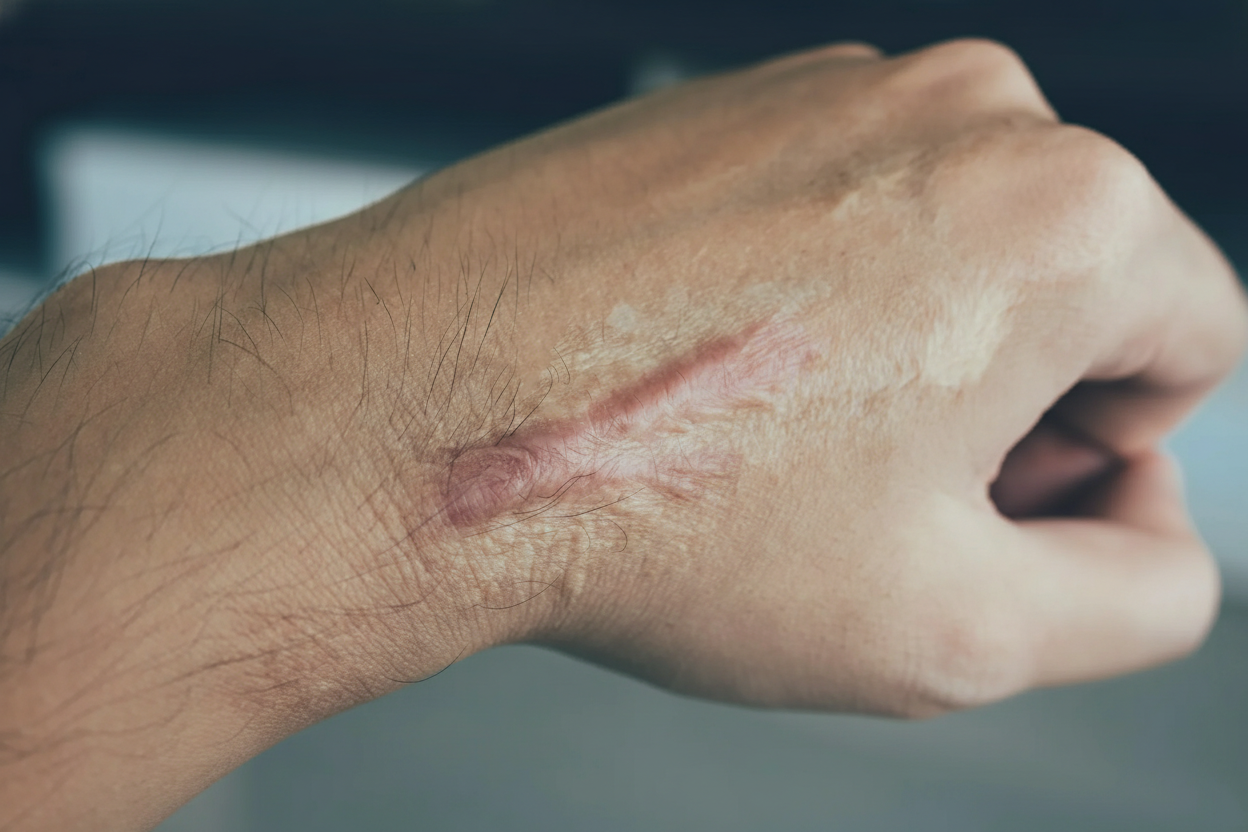
A 28-year-old woman follows up at an outpatient surgery clinic with an abnormal scarring of her incisional wound from an abdominal surgical procedure 6 months ago. She gives a history of a wound infection with a purulent discharge 1 week after surgery. On examination of the scar, a dense, raised, healed lesion is noted at the incision site. She also complains of an occasional itching sensation over the scar. There is no history of such scar changes in her family. An image of the lesion is given below. Which of the following statements best describe the scar abnormality?
A 4-year-old boy is brought to the emergency department for a right ankle injury sustained during a fall earlier that morning. His parents report that he is 'clumsy' when he runs and has fallen multiple times in the last year. He has reached most of his developmental milestones but did not walk until the age of 17 months. He is an only child and was adopted at age 1. He appears tearful and in mild distress. His temperature is 37.2°C (98.9°F), pulse is 72/min, respirations are 17/min, and blood pressure is 80/50 mm Hg. His right ankle is mildly swollen with no tenderness over the medial or lateral malleolus; range of motion is full with mild pain. He has marked enlargement of both calves. Patellar and Achilles reflexes are 1+ bilaterally. Strength is 4/5 in the deltoids, knee flexors/extensors, and 5/5 in the biceps and triceps. Babinski sign is absent. When standing up from a lying position, the patient crawls onto his knees and slowly walks himself up with his hands. Which of the following is the most likely underlying mechanism of this patient's condition?
A 21-year-old man comes to the military base physician for evaluation of progressive discomfort in his right shoulder for the past 4 months. He joined the military 6 months ago and is part of a drill team. In anticipation of an upcoming competition, he has been practicing rifle drills and firing exercises 8 hours a day. Physical examination shows tenderness to palpation and a firm mass in the superior part of the right deltopectoral groove. Range of motion is limited by pain and stiffness. Which of the following is the most likely diagnosis?
A 62-year-old woman comes to the physician because of worsening mental status over the past month. Her husband reports that she was initially experiencing lapses in memory but has recently started having difficulties performing activities of daily living. She appears withdrawn and avoids eye contact. Examination shows diffuse involuntary muscle jerking that can be provoked by loud noises. A cerebrospinal fluid analysis shows elevated concentration of 14-3-3 protein. Four months later, the patient dies. Pathologic examination of the brain on autopsy is most likely to show which of the following findings?
Practice by Chapter
Reversible cell injury mechanisms
Practice Questions
Irreversible cell injury (necrosis)
Practice Questions
Types of necrosis (coagulative, liquefactive, etc.)
Practice Questions
Apoptosis pathways
Practice Questions
Autophagy mechanisms
Practice Questions
Cellular adaptations (atrophy, hypertrophy)
Practice Questions
Hyperplasia and metaplasia
Practice Questions
Dysplasia
Practice Questions
Intracellular accumulations
Practice Questions
Pathologic calcification
Practice Questions
Cellular aging mechanisms
Practice Questions
Ischemia-reperfusion injury
Practice Questions
Free radical injury
Practice Questions
Want unlimited practice?
Get full access to all questions, explanations, and performance tracking.
Scan to download app