
Cardiovascular — MCQs

On this page
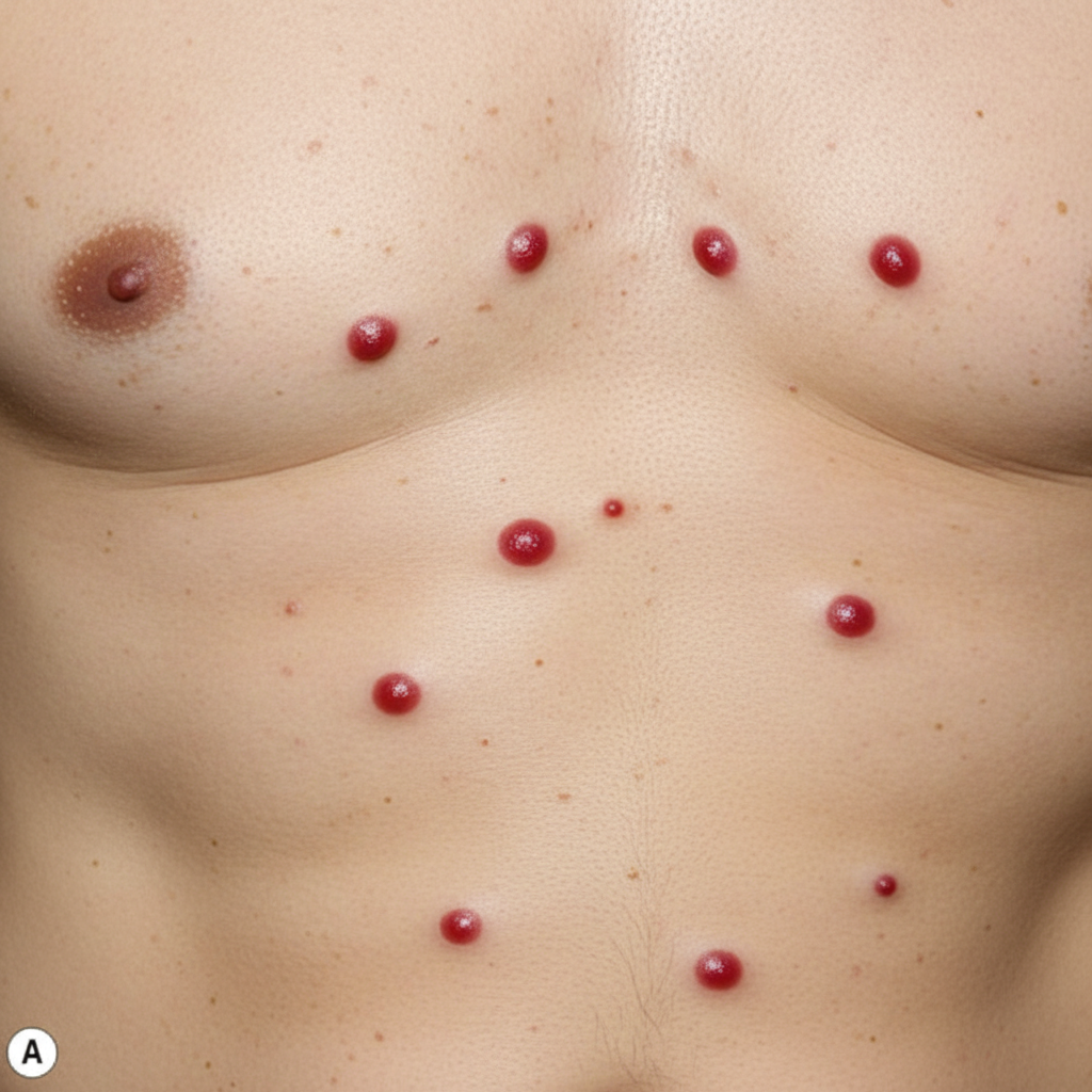
A 62-year-old man comes to the physician for evaluation of multiple red spots on his trunk. He first noticed these several months ago, and some appear to have increased in size. One day ago, he scratched one of these spots, and it bled for several minutes. Physical examination shows the findings in the photograph. Which of the following is the most likely diagnosis?
A 53-year-old man presents to the emergency department with a complaint of chest pain for 5 hours. The chest pain is continuous and squeezing in nature, not relieved by aspirin, and not related to the position of respiration. The blood pressure was 102/64 mm Hg, and the heart rate was 73/min. On physical examination, heart sounds are normal on auscultation. His ECG shows sinus rhythm with ST-segment elevation in leads II and III, aVF, and reciprocal segment depression in precordial leads V1–V6. Tissue plasminogen activator therapy is administered to the patient intravenously within 1 hour of arrival at the hospital. After 6 hours of therapy, the patient’s clinical condition starts to deteriorate. An ECG now shows ventricular fibrillation. The patient dies, despite all the efforts made in the intensive care unit. What is the most likely pathological finding to be expected in his heart muscles on autopsy?
A 59-year-old presents with right-sided hemiparesis, right-sided sensory loss, leftward eye deviation, and slurred speech. A head CT is performed which is significant for a hyperdense lesion affecting the putamen. The patient has a history of hypertension treated with hydrochlorothiazide, but is non-adherent. Which of the following is most likely associated with the cause of this patient’s neurological deficits?
A 63-year-old man presents to the emergency room with severe upper abdominal pain. His symptoms started 2 days prior to presentation and have progressed rapidly. He has been seen in the emergency room 3 times in the past year for acute alcohol intoxication. His past medical history is notable for multiple deep venous thromboses, hypertension, diabetes mellitus, gout, and a transient ischemic attack one year prior. He takes warfarin, lisinopril, metformin, glyburide, and allopurinol. His temperature is 100.0°F (37.8°C), blood pressure is 100/55 mmHg, pulse is 130/min, and respirations are 26/min. On exam, he is in acute distress but is able to answer questions appropriately. Hepatomegaly, splenomegaly, and scleral icterus are noted. There is a positive fluid wave. Laboratory analysis reveals an INR of 1.3. An abdominal ultrasound is ordered, and the patient is started on the appropriate management. However, before the ultrasound can begin, he rapidly loses consciousness and becomes unresponsive. He expires despite appropriate management. An autopsy the following day determines the cause of death to be a massive cerebrovascular accident. A liver biopsy demonstrates darkly erythematous congested areas in the centrilobular regions. This patient’s presenting symptoms are most likely caused by obstructive blood flow in which of the following vessels?
A 14-year-old girl is brought to the physician because of a 1-week history of malaise and chest pain. Three weeks ago, she had a sore throat that resolved without treatment. Her temperature is 38.7°C (101.7°F). Examination shows several subcutaneous nodules on her elbows and wrist bilaterally and a new-onset early systolic murmur best heard at the apex in the left lateral position. An endomysial biopsy is most likely to show which of the following?
A 51-year-old African American man with a history of poorly controlled hypertension presents to the emergency room with blurry vision and dyspnea. He reports rapid-onset blurred vision and difficulty breathing 4 hours prior to presentation. He takes lisinopril, hydrochlorothiazide, and spironolactone but has a history of poor medication compliance. He has a 50 pack-year smoking history and drinks 4-6 shots of vodka per day. His temperature is 99.2°F (37.3°C), blood pressure is 195/115 mmHg, pulse is 85/min, and respirations are 20/min. On exam, he is ill-appearing and pale. He is intermittently responsive and oriented to person but not place or time. Fundoscopic examination reveals swelling of the optic disc with blurred margins. A biopsy of this patient’s kidney would most likely reveal which of the following?
A 16-year-old boy is brought to the emergency department 20 minutes after collapsing while playing basketball. There is no personal or family history of serious illness. On arrival, there is no palpable pulse and no respiratory effort is seen. He is declared dead. The family agrees to an autopsy. Which of the following is most likely to be found in this patient?
A 22-year-old male varsity athlete visits the on-campus health services for shortness of breath, fatigue, and lower limb edema with onset 1 week after mild upper respiratory tract infection. Upon physical examination, his blood pressure is 100/68 mm Hg, heart rate is 120/min, respiratory rate is 23/min, and temperature is 36.4°C (97.5°F). He is referred to the nearest hospital, where his systolic pressure drops below 90 mm Hg with an S3 gallop, and he needs inotropic support in the critical care unit. A chest radiograph shows an enlarged heart, clear lungs, and effacement of the right costodiaphragmatic angle. A subsequent esophageal echocardiogram reveals severe dilation of all heart cavities, an ejection fraction of 23%, and mitral regurgitation. His family and personal history are unremarkable; therefore, an endomyocardial biopsy (EMB) is ordered. Which of the following microscopic findings would you expect in this specimen?
A 47-year-old man presents as a new patient at an outpatient clinic. He has never seen a physician before, but was motivated by his 40-year-old brother's recent heart attack and seeks to optimize his health. In particular, he read that uncontrolled atherosclerosis can lead to a heart attack. Which molecule is downregulated in response to the advent of atherosclerosis?
A 20-year-old Caucasian male presents with recurrent nosebleeds. Complete history reveals his father died in his 40's after an intracranial hemorrhage and two of his father's five siblings have also had recurrent nosebleeds. Which of the following would you expect to find in this patient?
Practice by Chapter
Atherosclerosis
Practice Questions
Ischemic heart disease
Practice Questions
Myocardial infarction pathology
Practice Questions
Hypertensive heart disease
Practice Questions
Congenital heart defects
Practice Questions
Valvular heart disease
Practice Questions
Rheumatic heart disease
Practice Questions
Infective endocarditis
Practice Questions
Myocarditis and cardiomyopathies
Practice Questions
Pericardial diseases
Practice Questions
Aneurysms and dissections
Practice Questions
Vasculitis
Practice Questions
Cardiac tumors
Practice Questions
Want unlimited practice?
Get full access to all questions, explanations, and performance tracking.
Scan to download app